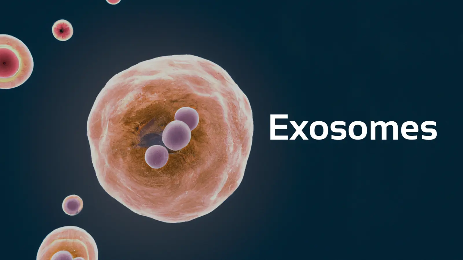
{"id":612,"date":"2026-07-23T10:05:17","date_gmt":"2026-07-23T09:05:17","guid":{"rendered":"https:\/\/ninaveli.com\/knowledge-hub\/?p=612"},"modified":"2026-07-23T10:05:17","modified_gmt":"2026-07-23T09:05:17","slug":"cellular-reprogramming-how-exosomes-and-paracrine-signaling-reset-the-regenerative-clock-in-aging-skin","status":"publish","type":"post","link":"https:\/\/ninaveli.com\/knowledge-hub\/cellular-reprogramming-how-exosomes-and-paracrine-signaling-reset-the-regenerative-clock-in-aging-skin\/","title":{"rendered":"Cellular Reprogramming: How Exosomes and Paracrine Signaling Reset the Regenerative Clock in Aging Skin"},"content":{"rendered":"\n<p class=\"wp-block-paragraph\">For years, stem cell therapy was hailed as the holy grail of regenerative medicine. Early protocols assumed that introducing stem cells into damaged tissue would allow those cells to engraft, differentiate, and replace old, dying skin structures.<\/p>\n\n\n\n<p class=\"wp-block-paragraph\">However, advanced molecular biology has revealed a fascinating biological truth: stem cells rarely engraft or replace tissue directly. Instead, their therapeutic power lies almost entirely in <strong>paracrine signaling<\/strong>\u2014the secretion of microscopic, message-carrying extracellular vesicles known as <strong>Exosomes<\/strong>.<\/p>\n\n\n\n<p class=\"wp-block-paragraph\">In aging, sun-damaged, or post-procedural skin, local dermal fibroblasts enter a state of metabolic fatigue or senescence, producing lower levels of collagen while secreting inflammatory enzymes.<\/p>\n\n\n\n<p class=\"wp-block-paragraph\">To reprogram these sluggish cells without invasive cellular transplants, pioneering biotech compaies such as Sinclair Pharma, <a href=\"https:\/\/ninaveli.com\/about.php\">Ninaveli<\/a>, and Evolus utilize <strong>Bio-Engineered Exosomes<\/strong>. These nano-vesicles deliver genetic instructions directly into target skin cells, turning back the cellular clock and accelerating tissue repair.<\/p>\n\n\n\n<h2 class=\"wp-block-heading\">1. The Paracrine Deficit: Why Aging Skin Loses Cellular Communication<\/h2>\n\n\n\n<p class=\"wp-block-paragraph\">Intercellular communication is the foundation of skin health. Young skin maintains an active network where cells constantly exchange signaling molecules to coordinate matrix repair and immune defense:<\/p>\n\n\n\n<ul class=\"wp-block-list\">\n<li><strong>Communication Breakdown:<\/strong> As skin ages, the quantity and functional quality of endogenously secreted extracellular vesicles decline precipitously.<\/li>\n\n\n\n<li><strong>Paracrine Isolation:<\/strong> Dermal fibroblasts become isolated from necessary regenerative signals. Lacking regulatory microRNA and growth factor triggers, fibroblasts downregulate Type I and Type III collagen synthesis.<\/li>\n\n\n\n<li><strong>Post-Traumatic Inflammatory Delay:<\/strong> Following aggressive aesthetic procedures (such as fractional CO<sub>2<\/sub> lasers or deep microneedling), aged or compromised skin struggles to transition from the initial inflammatory phase into the proliferative repair phase. This results in prolonged erythema, delayed re-epithelialization, and heightened risk of post-inflammatory hyperpigmentation (PIH).<\/li>\n<\/ul>\n\n\n\n<h2 class=\"wp-block-heading\">2. The Bio-Vesicular Cargo: How Exosomes Reprogram Target Cells<\/h2>\n\n\n\n<p class=\"wp-block-paragraph\"><strong>Exosomes<\/strong> are nanoscale extracellular vesicles (measuring 30 to 150 nanometers in diameter) bounded by a lipid bilayer membrane identical to human cell membranes.<\/p>\n\n\n\n<p class=\"wp-block-paragraph\">Rather than acting as simple single-target chemical actives, exosomes serve as complex <strong>biological delivery vehicles<\/strong> packed with thousands of signaling molecules:<\/p>\n\n\n\n<h3 class=\"wp-block-heading\">A. Molecular Cargo Composition<\/h3>\n\n\n\n<p class=\"wp-block-paragraph\">Each exosome carries a protected payload of bioactive information:<\/p>\n\n\n\n<ul class=\"wp-block-list\">\n<li><strong>MicroRNAs (miRNAs):<\/strong> Non-coding RNA fragments (such as miR-21, miR-29, and miR-133) that enter recipient cell nuclei to epigenetically downregulate pro-inflammatory enzymes and turn on collagen gene transcription.<\/li>\n\n\n\n<li><strong>Growth Factors:<\/strong> High concentrations of Transformative Growth Factor-beta (TGF-beta3), Vascular Endothelial Growth Factor (VEGF), and Platelet-Derived Growth Factor (PDGF) that stimulate rapid cell proliferation and healthy vessel formation.<\/li>\n\n\n\n<li><strong>Structural Proteins &amp; Peptides:<\/strong> Bio-identical signaling proteins that reinforce cell membrane receptors and jumpstart metabolic repair.<\/li>\n<\/ul>\n\n\n\n<h3 class=\"wp-block-heading\">B. Cellular Fusion and Nuclear Reprogramming<\/h3>\n\n\n\n<p class=\"wp-block-paragraph\">Because exosome membranes are composed of a natural phospholipid bilayer, they bypass standard cellular barriers. Upon reaching recipient dermal fibroblasts or basal keratinocytes, exosomes fuse directly with the host cell membrane or enter via endocytosis.<\/p>\n\n\n\n<p class=\"wp-block-paragraph\">Once inside, they release their microRNA and growth factor payload straight into the cytoplasm. This payload overrides senescent or sluggish signals, <strong>reprogramming recipient fibroblasts to behave like younger, highly active cells<\/strong>.<\/p>\n\n\n\n<h2 class=\"wp-block-heading\">3. Clinical Proof: What the Science Confirms<\/h2>\n\n\n\n<h3 class=\"wp-block-heading\">1. Accelerated Post-Laser Recovery and Inflammatory Modulation<\/h3>\n\n\n\n<p class=\"wp-block-paragraph\">In a clinical trial published in <em>PMC<\/em>, <strong>&#8220;Exosomes Derived from Stem Cells in Regenerative Dermatology and Post-Procedure Recovery,&#8221;<\/strong> researchers evaluated the impact of topical exosome administration following fractional resurfacing procedures.<\/p>\n\n\n\n<p class=\"wp-block-paragraph\">The objective clinical data demonstrated a <strong>statistically significant reduction in post-procedure erythema, edema, and downtime<\/strong> compared to vehicle controls.<\/p>\n\n\n\n<p class=\"wp-block-paragraph\">Histological evaluations confirmed that exosome-treated skin exhibited <strong>accelerated re-epithelialization, enhanced collagen fiber alignment, and a rapid drop in pro-inflammatory interleukins (IL-1beta and IL-6)<\/strong> within 48 to 72 hours post-treatment.<\/p>\n\n\n\n<h3 class=\"wp-block-heading\">2. Epigenetic Rejuvenation and Fibroblast Collagen Upregulation<\/h3>\n\n\n\n<p class=\"wp-block-paragraph\">According to a breakthrough study published in <em>ACS Nano (PMC)<\/em>, <strong>&#8220;Extracellular Vesicles (Exosomes) Reprogram Aged Dermal Fibroblasts to Increase Collagen Synthesis,&#8221;<\/strong> researchers tested the capacity of stem-cell-derived exosomes to reverse cellular aging in human dermal skin models.<\/p>\n\n\n\n<p class=\"wp-block-paragraph\">The study proved that exosome uptake by aged dermal fibroblasts resulted in a <strong>multifold upregulation of Type I procollagen gene expression<\/strong> alongside a significant reduction in collagen-degrading MMP-1 enzymes. The researchers concluded that exosomes actively reverse cellular senescence phenotypes by transferring functional microRNAs directly into target cells.<\/p>\n\n\n\n<h2 class=\"wp-block-heading\">4. Practice Summary<\/h2>\n\n\n\n<p class=\"wp-block-paragraph\">Exosomes represent the cutting edge of regenerative aesthetic medicine, shifting treatment paradigms from simple active ingredient topicality to true paracrine cellular reprogramming. By delivering microRNAs, growth factors, and regulatory proteins directly into recipient fibroblasts, bio-engineered exosomes reset cellular signaling pathways. This allows aesthetic practitioners to calm post-procedure inflammation, accelerate tissue recovery, and restore youthful collagen synthesis.<\/p>\n\n\n\n<h3 class=\"wp-block-heading\">Study Citations &amp; References<\/h3>\n\n\n\n<ul class=\"wp-block-list\">\n<li><strong>Study 1 (Exosomes in Post-Procedure Recovery &amp; Regeneration):<\/strong> <em>Exosomes Derived from Stem Cells in Regenerative Dermatology.<\/em>URL: <a href=\"https:\/\/pmc.ncbi.nlm.nih.gov\/articles\/PMC8398991\/\" target=\"_blank\" rel=\"noreferrer noopener\">https:\/\/pmc.ncbi.nlm.nih.gov\/articles\/PMC8398991\/<\/a><\/li>\n\n\n\n<li><strong>Study 2 (Fibroblast Reprogramming &amp; Collagen Gene Expression):<\/strong> <em>Exosomes Reprogram Aged Dermal Fibroblasts to Increase Collagen Synthesis.<\/em>URL: <a href=\"https:\/\/pmc.ncbi.nlm.nih.gov\/articles\/PMC7607370\/\" target=\"_blank\" rel=\"noreferrer noopener\">https:\/\/pmc.ncbi.nlm.nih.gov\/articles\/PMC7607370\/<\/a><\/li>\n\n\n\n<li><strong>Study 3 (Paracrine Mechanisms of Extracellular Vesicles):<\/strong> <em>Mesenchymal Stem Cell-Derived Exosomes in Skin Wound Healing and Tissue Repair.<\/em>URL: <a href=\"https:\/\/pmc.ncbi.nlm.nih.gov\/articles\/PMC8983239\/\" target=\"_blank\" rel=\"noreferrer noopener\">https:\/\/pmc.ncbi.nlm.nih.gov\/articles\/PMC8983239\/<\/a><\/li>\n<\/ul>\n\n\n\n<h2 class=\"wp-block-heading\">Frequently Asked Questions<\/h2>\n\n\n<div class=\"wp-block-uagb-faq uagb-faq__outer-wrap uagb-block-1599251e uagb-faq-icon-row uagb-faq-layout-accordion uagb-faq-expand-first-true uagb-faq-inactive-other-true uagb-faq__wrap uagb-buttons-layout-wrap uagb-faq-equal-height     \" data-faqtoggle=\"true\" role=\"tablist\"><div class=\"wp-block-uagb-faq-child uagb-faq-child__outer-wrap uagb-faq-item uagb-block-d1f815f4 \" role=\"tab\" tabindex=\"0\"><div class=\"uagb-faq-questions-button uagb-faq-questions\">\t\t\t<span class=\"uagb-icon uagb-faq-icon-wrap\">\n\t\t\t\t\t\t\t\t<svg xmlns=\"https:\/\/www.w3.org\/2000\/svg\" viewBox= \"0 0 448 512\"><path d=\"M432 256c0 17.69-14.33 32.01-32 32.01H256v144c0 17.69-14.33 31.99-32 31.99s-32-14.3-32-31.99v-144H48c-17.67 0-32-14.32-32-32.01s14.33-31.99 32-31.99H192v-144c0-17.69 14.33-32.01 32-32.01s32 14.32 32 32.01v144h144C417.7 224 432 238.3 432 256z\"><\/path><\/svg>\n\t\t\t\t\t\t\t<\/span>\n\t\t\t\t\t\t<span class=\"uagb-icon-active uagb-faq-icon-wrap\">\n\t\t\t\t\t\t\t\t<svg xmlns=\"https:\/\/www.w3.org\/2000\/svg\" viewBox= \"0 0 448 512\"><path d=\"M400 288h-352c-17.69 0-32-14.32-32-32.01s14.31-31.99 32-31.99h352c17.69 0 32 14.3 32 31.99S417.7 288 400 288z\"><\/path><\/svg>\n\t\t\t\t\t\t\t<\/span>\n\t\t\t<span class=\"uagb-question\">How do Exosomes differ from traditional Growth Factor serums or PRP (Platelet-Rich Plasma)?<\/span><\/div><div class=\"uagb-faq-content\"><p>Traditional growth factor serums contain free proteins that can degrade quickly on the skin surface and struggle to penetrate cell membranes due to their molecular size. PRP relies on the patient&#8217;s own blood platelets, meaning the quality and concentration of growth factors fluctuate based on the patient&#8217;s age, health, and lifestyle. <strong>Exosomes<\/strong> are pure, standardized nano-vesicles that protect their internal payload of microRNAs and growth factors inside a protective lipid bilayer. This membrane allows them to fuse directly with target skin cell membranes, delivering precise genetic instructions without relying on the patient&#8217;s age or systemic health.<\/p><\/div><\/div><div class=\"wp-block-uagb-faq-child uagb-faq-child__outer-wrap uagb-faq-item uagb-block-42d2472e \" role=\"tab\" tabindex=\"0\"><div class=\"uagb-faq-questions-button uagb-faq-questions\">\t\t\t<span class=\"uagb-icon uagb-faq-icon-wrap\">\n\t\t\t\t\t\t\t\t<svg xmlns=\"https:\/\/www.w3.org\/2000\/svg\" viewBox= \"0 0 448 512\"><path d=\"M432 256c0 17.69-14.33 32.01-32 32.01H256v144c0 17.69-14.33 31.99-32 31.99s-32-14.3-32-31.99v-144H48c-17.67 0-32-14.32-32-32.01s14.33-31.99 32-31.99H192v-144c0-17.69 14.33-32.01 32-32.01s32 14.32 32 32.01v144h144C417.7 224 432 238.3 432 256z\"><\/path><\/svg>\n\t\t\t\t\t\t\t<\/span>\n\t\t\t\t\t\t<span class=\"uagb-icon-active uagb-faq-icon-wrap\">\n\t\t\t\t\t\t\t\t<svg xmlns=\"https:\/\/www.w3.org\/2000\/svg\" viewBox= \"0 0 448 512\"><path d=\"M400 288h-352c-17.69 0-32-14.32-32-32.01s14.31-31.99 32-31.99h352c17.69 0 32 14.3 32 31.99S417.7 288 400 288z\"><\/path><\/svg>\n\t\t\t\t\t\t\t<\/span>\n\t\t\t<span class=\"uagb-question\">Can Exosomes be used immediately following energy-based treatments like RF microneedling or fractional lasers?<\/span><\/div><div class=\"uagb-faq-content\"><p>Yes, applying Exosomes immediately post-treatment is one of their primary clinical applications. Procedures like RF microneedling, fractional $CO_2$ lasers, and chemical peels create controlled micro-channels in the skin barrier. Applying an Exosome formulation through these open channels allows nano-vesicles to penetrate directly into the papillary dermis, where they calm post-procedure redness, suppress inflammatory cytokines, and accelerate re-epithelialization.<\/p><\/div><\/div><div class=\"wp-block-uagb-faq-child uagb-faq-child__outer-wrap uagb-faq-item uagb-block-4191d11e \" role=\"tab\" tabindex=\"0\"><div class=\"uagb-faq-questions-button uagb-faq-questions\">\t\t\t<span class=\"uagb-icon uagb-faq-icon-wrap\">\n\t\t\t\t\t\t\t\t<svg xmlns=\"https:\/\/www.w3.org\/2000\/svg\" viewBox= \"0 0 448 512\"><path d=\"M432 256c0 17.69-14.33 32.01-32 32.01H256v144c0 17.69-14.33 31.99-32 31.99s-32-14.3-32-31.99v-144H48c-17.67 0-32-14.32-32-32.01s14.33-31.99 32-31.99H192v-144c0-17.69 14.33-32.01 32-32.01s32 14.32 32 32.01v144h144C417.7 224 432 238.3 432 256z\"><\/path><\/svg>\n\t\t\t\t\t\t\t<\/span>\n\t\t\t\t\t\t<span class=\"uagb-icon-active uagb-faq-icon-wrap\">\n\t\t\t\t\t\t\t\t<svg xmlns=\"https:\/\/www.w3.org\/2000\/svg\" viewBox= \"0 0 448 512\"><path d=\"M400 288h-352c-17.69 0-32-14.32-32-32.01s14.31-31.99 32-31.99h352c17.69 0 32 14.3 32 31.99S417.7 288 400 288z\"><\/path><\/svg>\n\t\t\t\t\t\t\t<\/span>\n\t\t\t<span class=\"uagb-question\">How long does it take to see visible skin quality improvements following an Exosome protocol?<\/span><\/div><div class=\"uagb-faq-content\"><p><br>Due to their rapid anti-inflammatory and cellular repair signaling, initial post-procedure benefits\u2014such as reduced redness, calmed irritation, and improved skin smoothness\u2014are frequently visible within <strong>24 to 72 hours<\/strong>. Longer-term structural benefits\u2014including increased collagen density, improved skin tightness, and refined texture\u2014develop over <strong>3 to 6 weeks<\/strong> as reprogrammed fibroblasts synthesize new extracellular matrix components.<\/p><\/div><\/div><\/div>\n\n\n<p class=\"wp-block-paragraph\"><strong><em>Disclaimer:<\/em><\/strong><em>\u00a0The content provided in the Ninaveli Knowledge Hub is for informational and educational purposes only. This content is not intended to be a substitute for professional medical advice, diagnosis, or treatment and should not be used as such. Always seek the advice of a qualified healthcare provider or dermatologist with any questions you may have regarding a medical condition or before starting any new skincare regimen. Ninaveli does not guarantee the accuracy, completeness, or timeliness of the information provided and assumes no liability for any actions taken based on this content.<\/em><\/p>\n","protected":false},"excerpt":{"rendered":"<p>For years, stem cell therapy was hailed as the holy grail of regenerative medicine. Early protocols assumed that introducing stem cells into damaged tissue would allow those cells to engraft, differentiate, and replace old, dying skin structures. However, advanced molecular biology has revealed a fascinating biological truth: stem cells rarely engraft or replace tissue directly. [&hellip;]<\/p>\n","protected":false},"author":1,"featured_media":613,"comment_status":"closed","ping_status":"","sticky":false,"template":"","format":"standard","meta":{"_uag_custom_page_level_css":"","_swt_meta_header_display":false,"_swt_meta_footer_display":false,"_swt_meta_site_title_display":false,"_swt_meta_sticky_header":false,"_swt_meta_transparent_header":false,"footnotes":""},"categories":[7],"tags":[],"class_list":["post-612","post","type-post","status-publish","format-standard","has-post-thumbnail","hentry","category-articles"],"uagb_featured_image_src":{"full":["https:\/\/ninaveli.com\/knowledge-hub\/wp-content\/uploads\/2026\/07\/exosomes.jpg",1489,992,false],"thumbnail":["https:\/\/ninaveli.com\/knowledge-hub\/wp-content\/uploads\/2026\/07\/exosomes-150x150.jpg",150,150,true],"medium":["https:\/\/ninaveli.com\/knowledge-hub\/wp-content\/uploads\/2026\/07\/exosomes-300x200.jpg",300,200,true],"medium_large":["https:\/\/ninaveli.com\/knowledge-hub\/wp-content\/uploads\/2026\/07\/exosomes-768x512.jpg",768,512,true],"large":["https:\/\/ninaveli.com\/knowledge-hub\/wp-content\/uploads\/2026\/07\/exosomes-1024x682.jpg",1024,682,true],"1536x1536":["https:\/\/ninaveli.com\/knowledge-hub\/wp-content\/uploads\/2026\/07\/exosomes.jpg",1489,992,false],"2048x2048":["https:\/\/ninaveli.com\/knowledge-hub\/wp-content\/uploads\/2026\/07\/exosomes.jpg",1489,992,false]},"uagb_author_info":{"display_name":"Ninaveli","author_link":"https:\/\/ninaveli.com\/knowledge-hub\/author\/daniel\/"},"uagb_comment_info":0,"uagb_excerpt":"For years, stem cell therapy was hailed as the holy grail of regenerative medicine. Early protocols assumed that introducing stem cells into damaged tissue would allow those cells to engraft, differentiate, and replace old, dying skin structures. However, advanced molecular biology has revealed a fascinating biological truth: stem cells rarely engraft or replace tissue directly.&hellip;","_links":{"self":[{"href":"https:\/\/ninaveli.com\/knowledge-hub\/wp-json\/wp\/v2\/posts\/612","targetHints":{"allow":["GET"]}}],"collection":[{"href":"https:\/\/ninaveli.com\/knowledge-hub\/wp-json\/wp\/v2\/posts"}],"about":[{"href":"https:\/\/ninaveli.com\/knowledge-hub\/wp-json\/wp\/v2\/types\/post"}],"author":[{"embeddable":true,"href":"https:\/\/ninaveli.com\/knowledge-hub\/wp-json\/wp\/v2\/users\/1"}],"replies":[{"embeddable":true,"href":"https:\/\/ninaveli.com\/knowledge-hub\/wp-json\/wp\/v2\/comments?post=612"}],"version-history":[{"count":1,"href":"https:\/\/ninaveli.com\/knowledge-hub\/wp-json\/wp\/v2\/posts\/612\/revisions"}],"predecessor-version":[{"id":614,"href":"https:\/\/ninaveli.com\/knowledge-hub\/wp-json\/wp\/v2\/posts\/612\/revisions\/614"}],"wp:featuredmedia":[{"embeddable":true,"href":"https:\/\/ninaveli.com\/knowledge-hub\/wp-json\/wp\/v2\/media\/613"}],"wp:attachment":[{"href":"https:\/\/ninaveli.com\/knowledge-hub\/wp-json\/wp\/v2\/media?parent=612"}],"wp:term":[{"taxonomy":"category","embeddable":true,"href":"https:\/\/ninaveli.com\/knowledge-hub\/wp-json\/wp\/v2\/categories?post=612"},{"taxonomy":"post_tag","embeddable":true,"href":"https:\/\/ninaveli.com\/knowledge-hub\/wp-json\/wp\/v2\/tags?post=612"}],"curies":[{"name":"wp","href":"https:\/\/api.w.org\/{rel}","templated":true}]}}