한국어
 Русский
 العربية
 עברית
 Français
 Deutsch
 Español
Re-Anchoring the Epidermis: How rhCollagen Restores the Dermal-Epidermal Junction
In the modern aesthetic industry, addressing facial sagging has long focused on the macro-layers: replacing subcutaneous fat loss with dermal fillers or tightening deep fascial layers with energy-based devices. However, a major architectural discovery has shifted focus to a microscopic, critical interface: the Dermal-Epidermal Junction (DEJ).
When patients complain of a loss of skin “firmness,” global sloughing, or tissue fragility, the primary defect is often located directly at this basement membrane boundary. As we age, the once wave-like, interlocking structure of the DEJ flattens out, causing the upper skin layer (epidermis) to lose its anchor to the deeper matrix (dermis).
To repair this critical anatomical bridge without relying on animal-derived proteins or foreign inflammatory gels, advanced aesthetic medicine is turning to Synthetic Recombinant Human Collagen (rhCollagen), a highly effective ingredient currently being invested in by companies like Meytox, Ninaveli, and Sinclair Pharma.
Here is a comprehensive breakdown of the DEJ, how its flattening drives facial sagging, and the cellular mechanics of how rhCollagen restores this foundational boundary.
The Target Concern: Dermal-Epidermal Junction (DEJ) Flattening & Structural Sagging
The Dermal-Epidermal Junction is a specialized, complex basement membrane zone that physically welds the epidermis to the underlying dermis.
In young, healthy skin, the DEJ is not a flat line; it is a series of finger-like interlocking projections known as rete ridges (extending down from the epidermis) and dermal papillae (extending up from the dermis). This undulating design serves two vital functions:
- Mechanical Interlocking: It creates a high surface area that physically resists shear stress and gravitational drag, preventing the epidermis from sliding over the dermis.
- Nutritional and Signal Transfer: The epidermis contains no blood vessels. It relies entirely on the capillary beds inside the dermal papillae across the DEJ for oxygen, glucose, and growth factor diffusion.
How It Forms: The Flattening Cascade
As intrinsic aging and chronic photo-damage progress, the mechanical architecture of the DEJ undergoes severe degradation:
- Anchoring Fibril Degradation: Type IV and Type VII collagen fibers—the structural “rivets” that anchor the lamina densa to the papillary dermis—are progressively degraded by overactive matrix metalloproteinase (MMP) enzymes.
- Rete Ridge Effacement: The undulating rete ridges flatten out. The surface area of the boundary collapses by up to 50% between youth and old age.
- Nutritional Starvation and Tissue Sliding: With a flattened DEJ, the exchange of nutrients drops dramatically. Epidermal stem cell proliferation slows, the epidermis thins, and the skin loses its structural anchorage. Gravitational pull now causes the unanchored epidermis to slide downward, manifesting cosmetically as crepey, sagging, and structurally compromised skin.
Enter Recombinant Human Collagen (rhCollagen)
Historically, cosmetic formulations utilized animal-derived collagens (extracted from bovine or porcine sources). However, animal collagens carry inherent risks of immunogenicity, batch variability, and pathogen transmission, and their altered molecular structures often fail to integrate cleanly with human tissue.
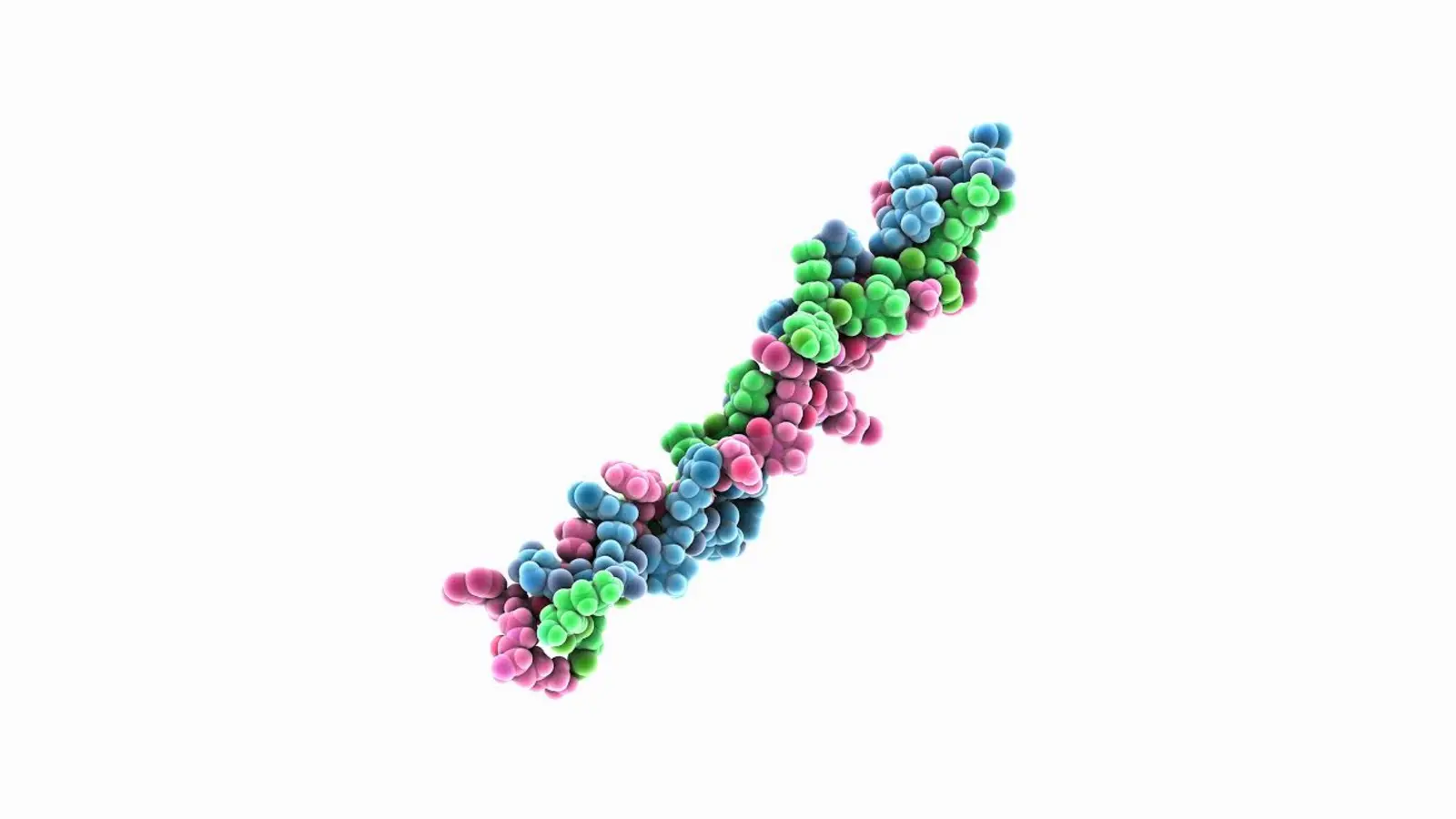
Recombinant Human Collagen (rhCollagen) represents a breakthrough in genetic bio-engineering. Expressed through bio-fermentation platforms (such as transgenic plants or specialized yeast vectors), rhCollagen matches the exact genetic sequence and triple-helical architecture of native human Type I and Type III collagen alpha chains.
The Cellular Mechanism: How It Restores the Boundary
When intradermally delivered into the upper papillary dermis and DEJ zone, rhCollagen bypasses foreign-body responses to act as a direct functional biomaterial.
First, rhCollagen Micro-Molecules Integrate Seamlessly into the extracellular matrix. Because its amino acid sequence is 100% human-identical, it exhibits zero immunogenic rejection, allowing local integrins on keratinocytes and fibroblasts to bind directly to the protein.
Next, this cellular binding triggers Integrin-Mediated Signal Transduction. Basal keratinocytes respond by upregulating the gene expression of essential basement membrane proteins, specifically Type IV Collagen, Type VII Collagen, and Laminin-511.
This surge in anchoring proteins initiates Basement Membrane Reconstruction. The new Type VII collagen molecules form stable anchoring fibrils that loop from the lamina densa deep into the papillary dermis. As the basement membrane rebuilds, basal keratinocytes increase their proliferation rate, reforming the physical finger-like Rete Ridges. The physical bond between the dermis and epidermis is re-anchored, pulling sagging surface tissue taut and restoring natural mechanical firmness.
According to a molecular study published in PMC, “Regeneration of collagen fibrils at the papillary dermis by reconstructing basement membrane at the dermal–epidermal junction,” structural reconstruction of the basement membrane complex directly stimulates epidermal keratinocytes to secrete PDGF-BB. This signaling surge triggers local dermal fibroblasts to synthesize fresh Type I and Type III procollagen fibrils directly beneath the junction, reinforcing the entire sub-epidermal layer.
Clinical Proof: What the Science Says
The application of recombinant human collagen variants and basement-membrane targeted protocols is validated by recent clinical literature.
1. Significant Improvement in Skin Texture and Firmness
According to a clinical study published in PMC, “Recombinant Type III Humanized Collagen Solution for Injection Promotes Skin Repair in Chinese Population: A Case Series,” rhCollagen administration delivers rapid structural repair.
The study evaluated subjects receiving intradermal rhCollagen injections. Quantitative objective imaging (VISIA) confirmed a statistically significant improvement in skin firmness, elasticity, and surface smoothness within 14 to 30 days post-baseline. Evaluators observed that the bio-identical protein reduced signs of skin laxity and tissue fragility without triggering post-treatment granulomas or persistent inflammation.
2. High Penetration and Fibroblast Proliferation Data
To evaluate the exact cellular interaction of recombinant human collagen, an in vitro and ex vivo study published in PMC, “Development of a highly effective recombinant protein from human collagen type III Alpha 1 (COL3A1) to enhance human skin cell functionality,” analyzed the biological impact of rhCOL3A1 fragments.
The study demonstrated that functional recombinant human collagen fragments specifically penetrate both the epidermal and dermal layers in full-thickness models and directly stimulate human dermal fibroblast proliferation. Furthermore, the recombinant protein significantly upregulates endogenous collagen biosynthesis, proving that rhCollagen functions as both an architectural scaffold and a bio-active cell stimulator.
The Takeaway for Practitioners and Patients
Recombinant Human Collagen (rhCollagen) represents a major step forward in precision regenerative aesthetics. By targeting the structural degradation of the Dermal-Epidermal Junction rather than simply placing volume underneath it, providers can address the root cause of surface laxity and crepey texture. Re-anchoring the basement membrane with human-identical proteins restores the mechanical bond between the skin layers, delivering a smooth, firm, and resilient complexion.
Study Citations & References
- Study 1 (Basement Membrane & DEJ Reconstruction): Regeneration of collagen fibrils at the papillary dermis by reconstructing basement membrane at the dermal–epidermal junction.URL: https://pmc.ncbi.nlm.nih.gov/articles/PMC8764085/
- Study 2 (Clinical rhCollagen Repair Data): Recombinant Type III Humanized Collagen Solution for Injection Promotes Skin Repair in Chinese Population: A Case Series.URL: https://pmc.ncbi.nlm.nih.gov/articles/PMC12104008/
- Study 3 (Fibroblast Proliferation & Penetration Data): Development of a highly effective recombinant protein from human collagen type III Alpha 1 (COL3A1) to enhance human skin cell functionality.URL: https://pmc.ncbi.nlm.nih.gov/articles/PMC11444990/
Frequently asked questions
How does Recombinant Human Collagen (rhCollagen) compare to traditional animal-derived collagen in aesthetic medicine?
The primary difference lies in immunogenicity, biological purity, and functional cellular integration. Traditional animal-derived collagens (extracted from bovine, porcine, or marine sources) carry a significant risk of triggering allergic reactions or foreign-body immune responses because their amino acid sequences differ from human genetics. Consequently, animal collagen often requires pre-treatment skin patch testing and can form rigid, non-physiological clumps in the tissue.
In contrast, rhCollagen is bio-engineered via genetic fermentation to match human Type I or Type III collagen sequences 100%. Because it is human-identical, it exhibits zero immunogenic rejection, carries no risk of animal-borne pathogen transmission, and integrates cleanly with native skin tissue. Instead of acting as an inert foreign implant, rhCollagen actively binds to human cell receptors (integrins) to stimulate genuine tissue regeneration.
Why is restoring the Dermal-Epidermal Junction (DEJ) more effective for crepey skin than traditional volume fillers?
Crepey skin and surface structural laxity are primarily driven by a flattening of the microscopic boundary connecting the top layer of skin (epidermis) to the deeper layer (dermis). Traditional dermal fillers (such as crosslinked hyaluronic acid) are designed to replace deep volumetric fat loss or lift heavy folds; when injected superficially to treat crepey texture, they can create unnatural ridge lumps or bluish shadows (the Tyndall effect).
Targeting the DEJ with rhCollagen restores the microscopic “anchoring fibrils” (Type IV and VII collagen) that weld the skin layers together. Re-anchoring the basement membrane restores the interlocking “rete ridges,” allowing nutrients to flow back into the epidermis and pulling the loose, crepey skin surface taut without adding artificial or unnatural volume.
How quickly do patients see structural improvements after an rhCollagen treatment protocol?
Because rhCollagen provides both instant structural support and long-term biological cell signaling, results develop in two distinct phases. Immediately post-treatment, patients notice an initial improvement in skin hydration and surface smoothness as the bio-identical protein matrix settles into the upper papillary dermis.
However, the primary regenerative outcome—the synthesis of new native anchoring fibrils and the restructuring of the DEJ—requires cellular activity. Clinical trials demonstrate that measurable increases in skin firmness, elasticity, and density typically peak between 14 to 30 days following the treatment session, with optimal tissue remodeling continuing over subsequent weeks as local fibroblasts proliferate.
Disclaimer: The content of the Ninaveli Knowledge Hub is for informational and educational purposes only. It is not a substitute for professional medical advice, diagnosis or treatment. Always seek the advice of a qualified healthcare provider with any questions you may have regarding a medical condition or before starting any new treatment. Ninaveli does not guarantee the accuracy, completeness or timeliness of the information and assumes no liability for any actions taken based on it.