

 한국어
한국어  Русский
  العربية
  עברית
עברית  Français
  Deutsch
  Español
Knowledge Hub
The science of beautiful skin, explained.
Ingredient breakdowns, clinical articles and the latest research on regenerative aesthetics, with references to the published studies.
66 articles
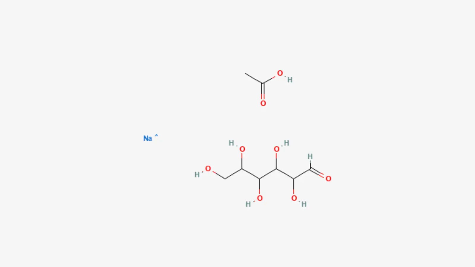
What is CMC: The Scientific Mechanisms and Clinical Evidence
Read article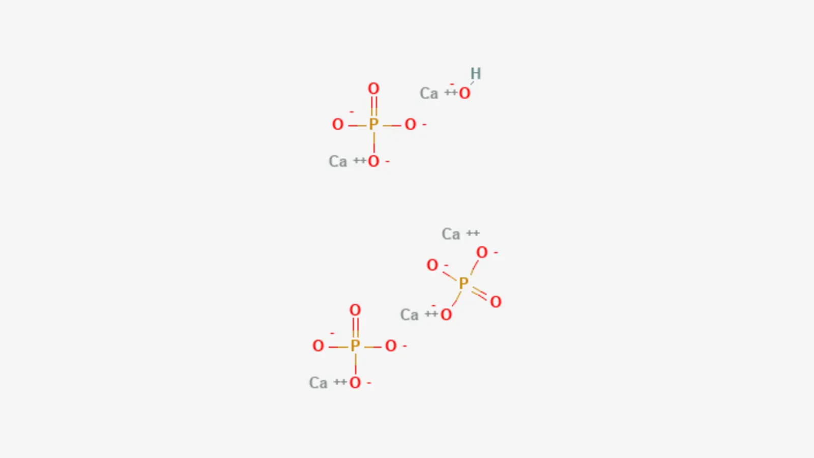
What is Calcium Hydroxylapatite: The Scientific Mechanisms and Clinical Evidence
Read article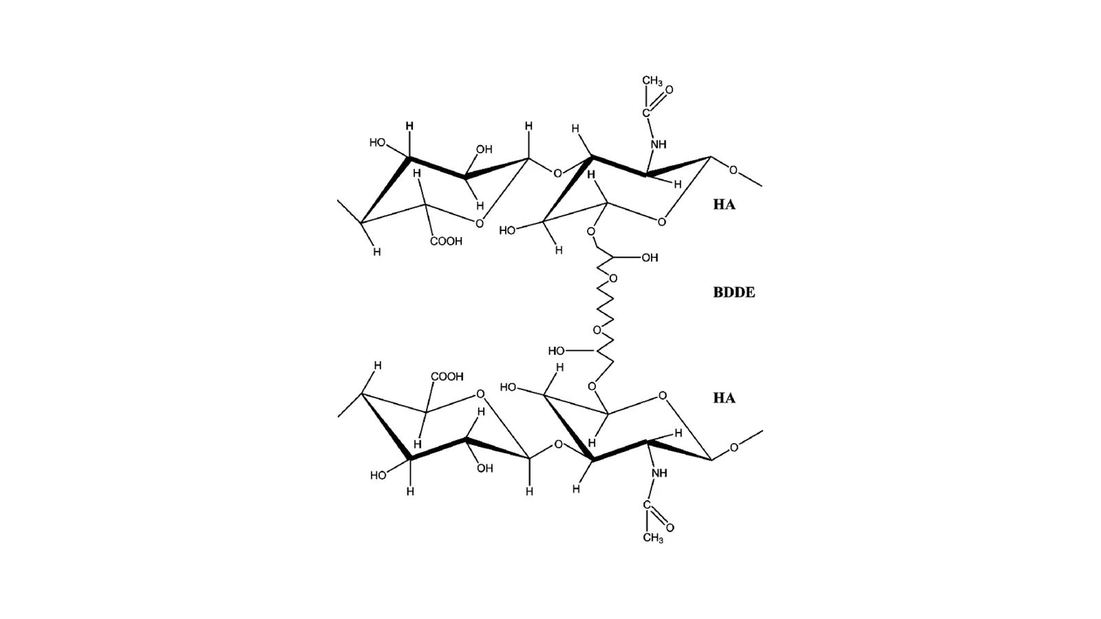
What is Crosslinked Hyaluronic Acid: The Scientific Mechanisms and Clinical Evidence
Read article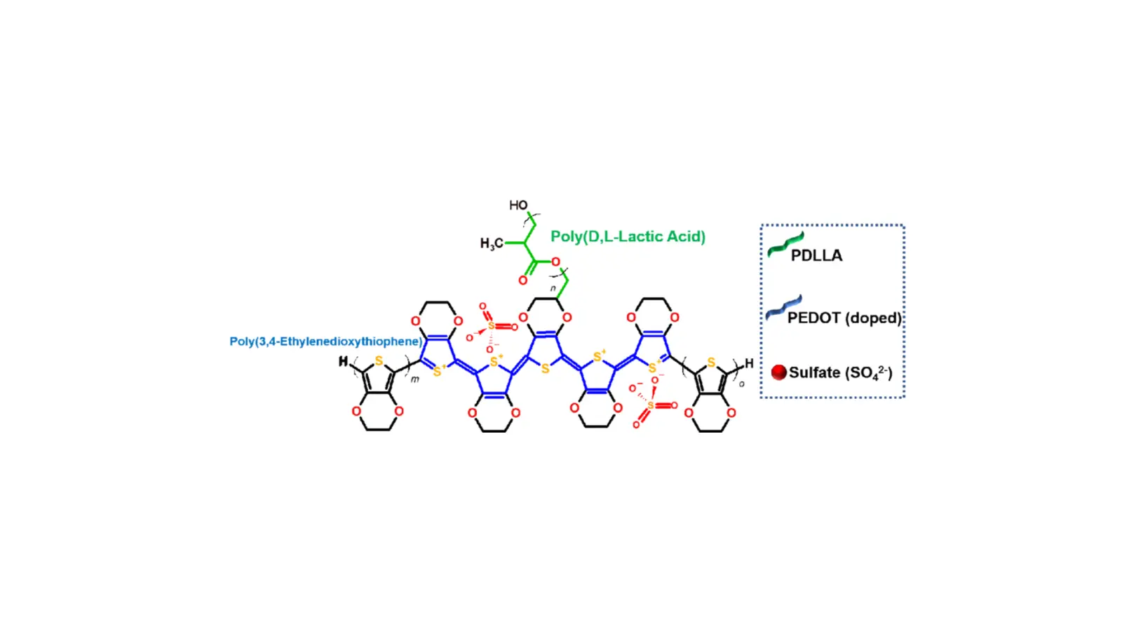
What is PDLLA: The Scientific Mechanisms and Clinical Evidence
Read article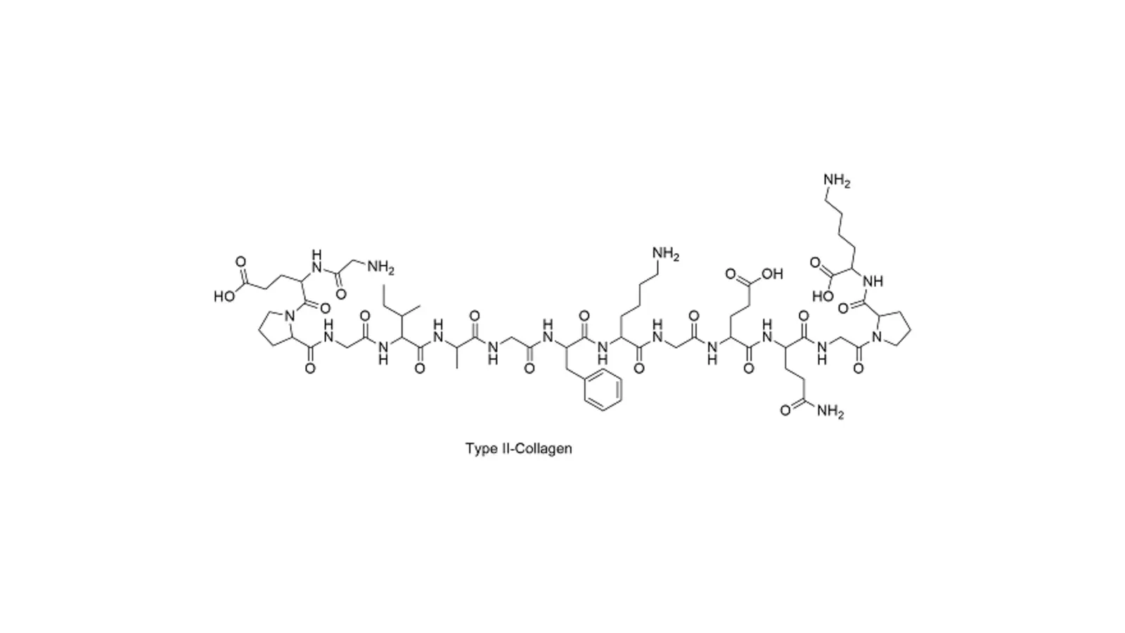
What is RH Collagen Type 1: The Scientific Mechanisms and Clinical Evidence
Read article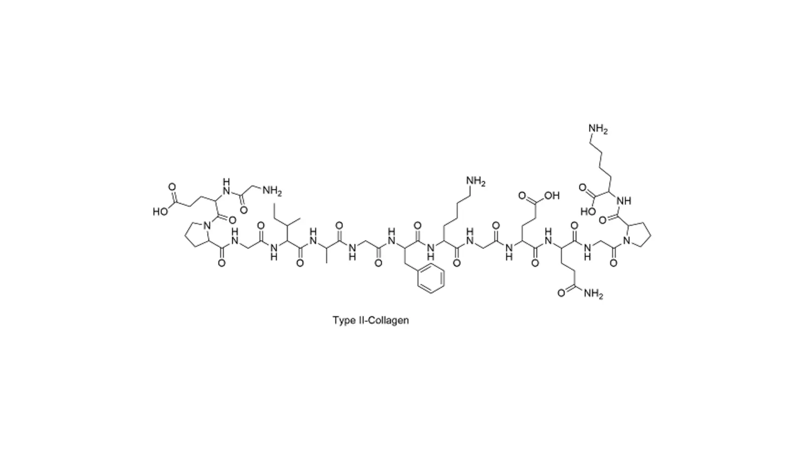
What is RH Collagen Type 3: The Scientific Mechanisms and Clinical Evidence
Read article
What are Peptides: The Scientific Mechanisms and Clinical Evidence
Read article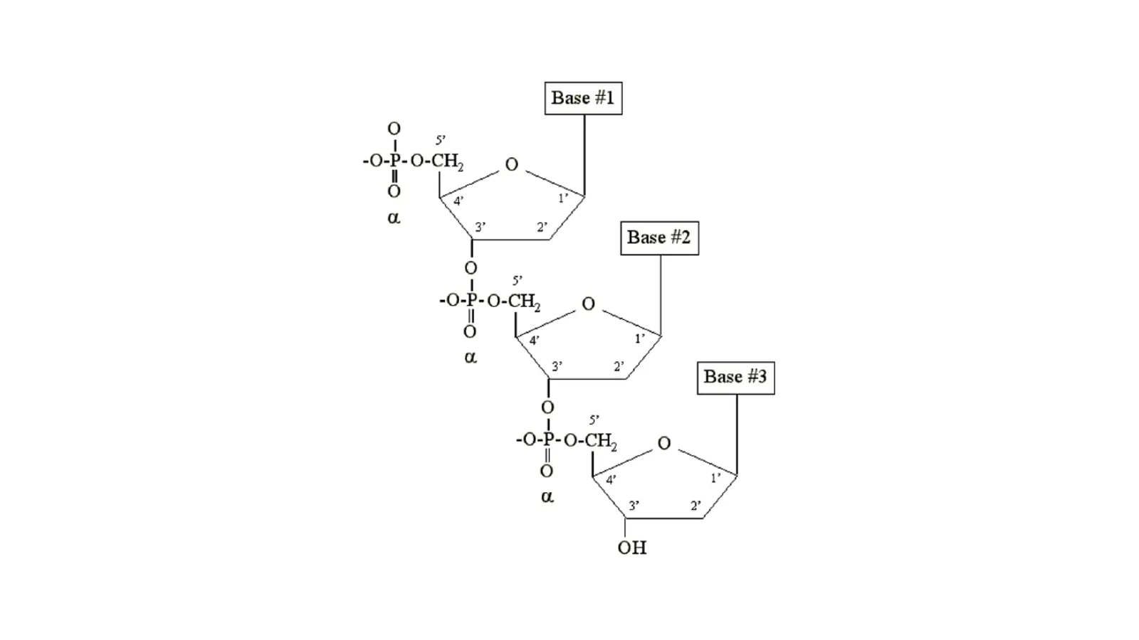
What are Polynucleotides: The Scientific Mechanisms and Clinical Evidence
Read article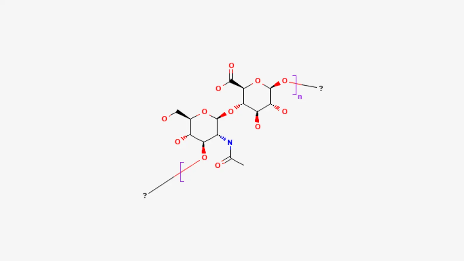
What is H-HA: The Scientific Mechanisms and Clinical Evidence
Read article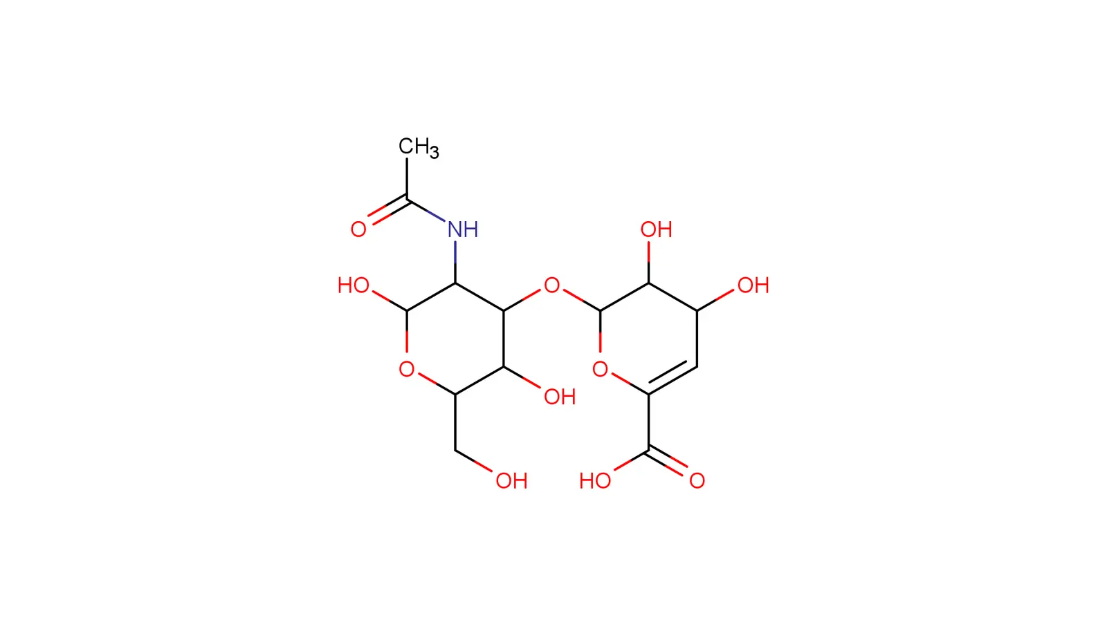
What is Hyaluronic Acid: The Scientific Mechanisms and Clinical Evidence
Read article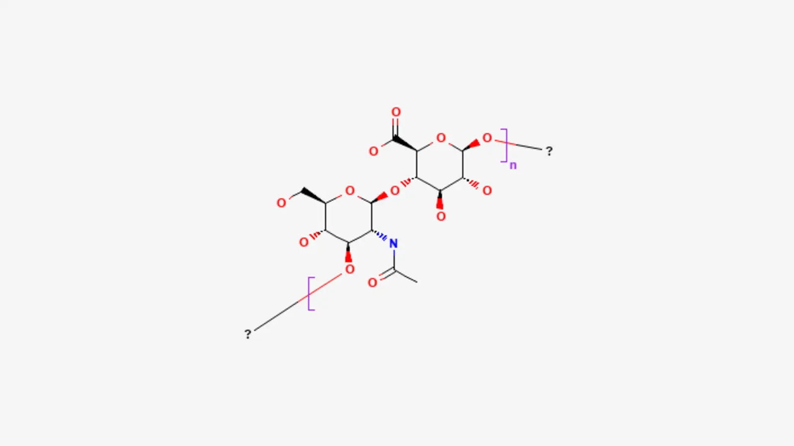
What is L-HA: The Scientific Mechanisms and Clinical Evidence
Read article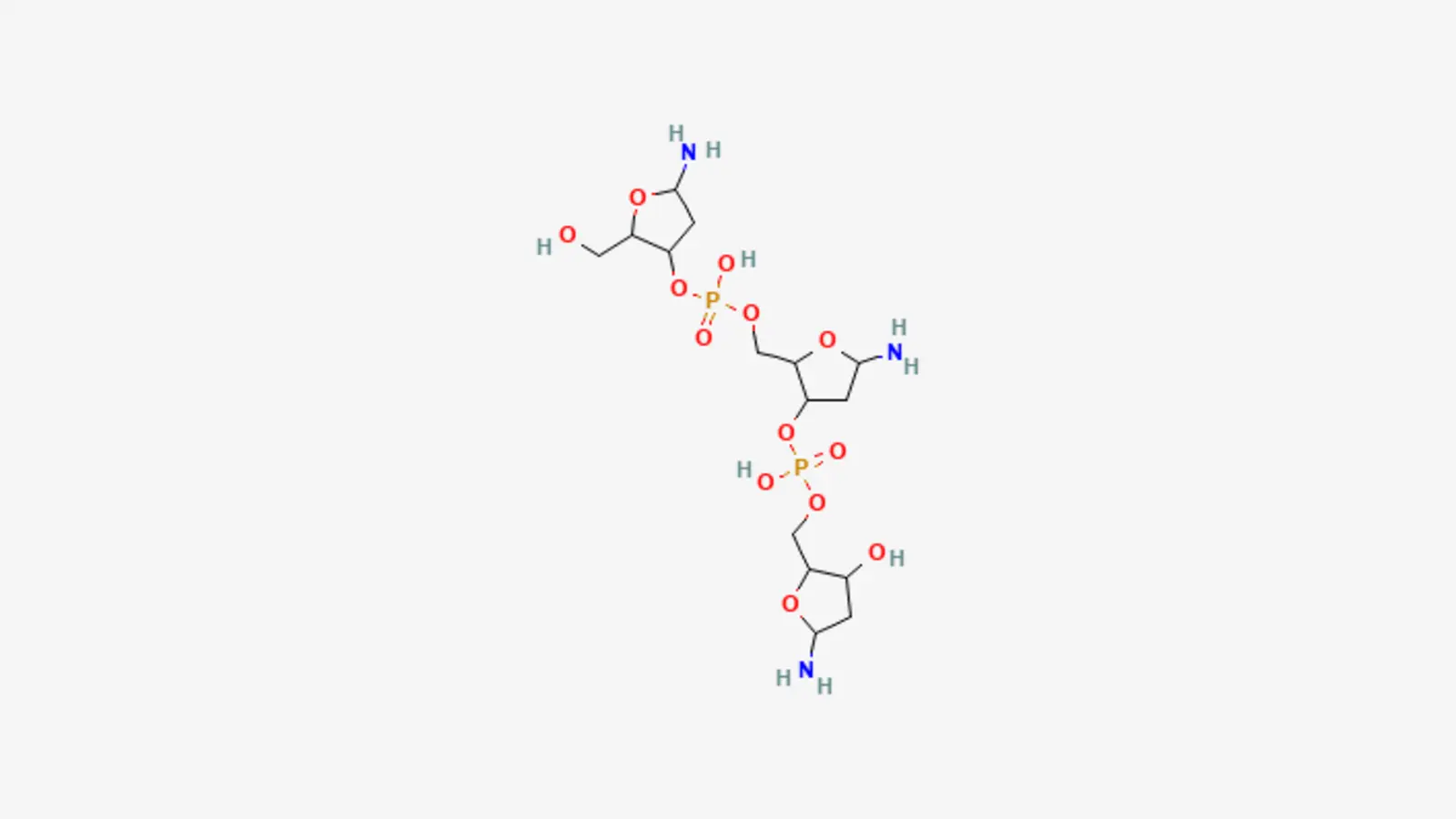
What is PDRN: The Scientific Mechanisms and Clinical Evidence
Read article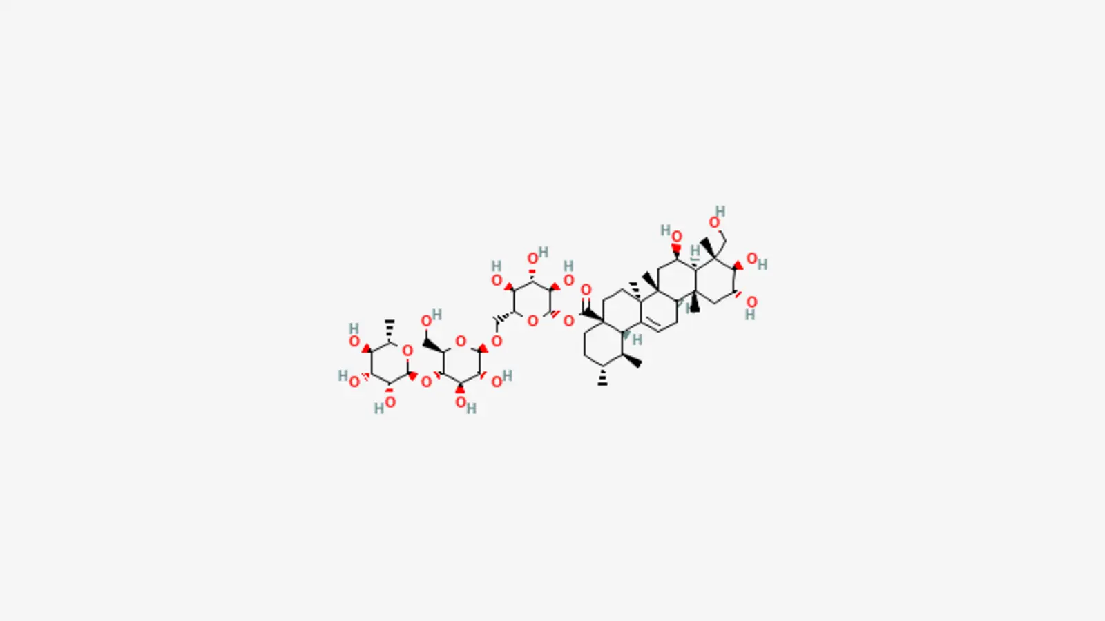
What is Cica: Scientific mechanisms and Clinical Evidence
Read article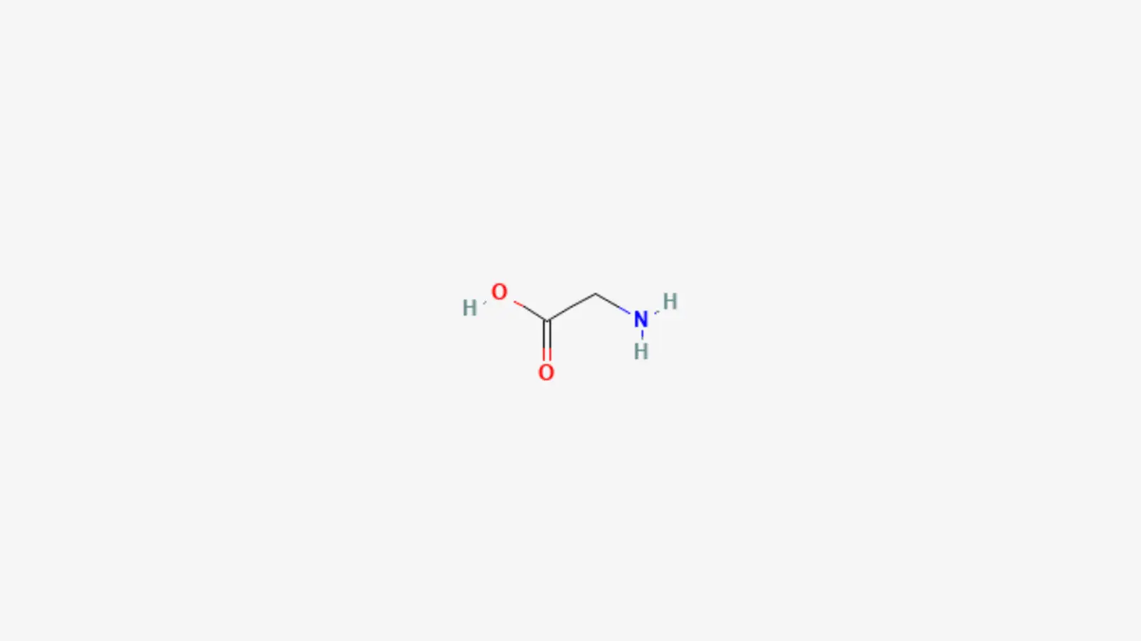
What is Glycine: Scientific Mechanisms and Clinical Evidence
Read article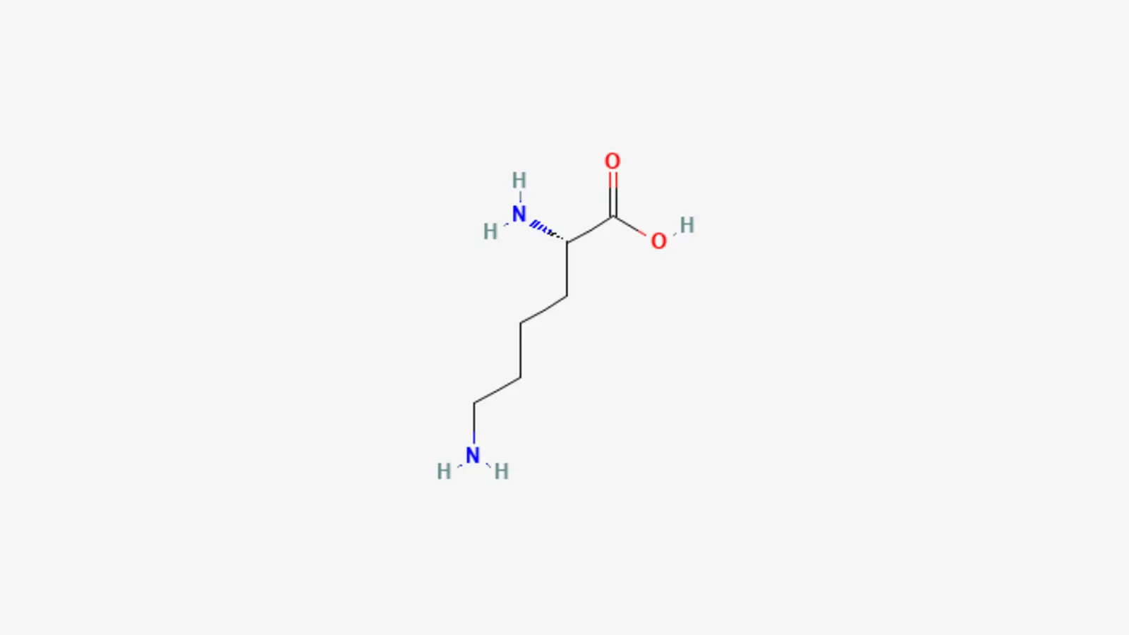
What is Lysine: Scientific Mechanisms and Clinical Evidence
Read article
What is Mannitol: The Scientific Mechanisms and Clinical Evidence
Read article
What is NMN: The Scientific Mechanisms and Clinical Evidence
Read article
What is NR: The Scientific Mechanisms and Clinical Evidence
Read article
What is Niacinamide: The Scientific Mechanisms and Clinical Evidence
Read article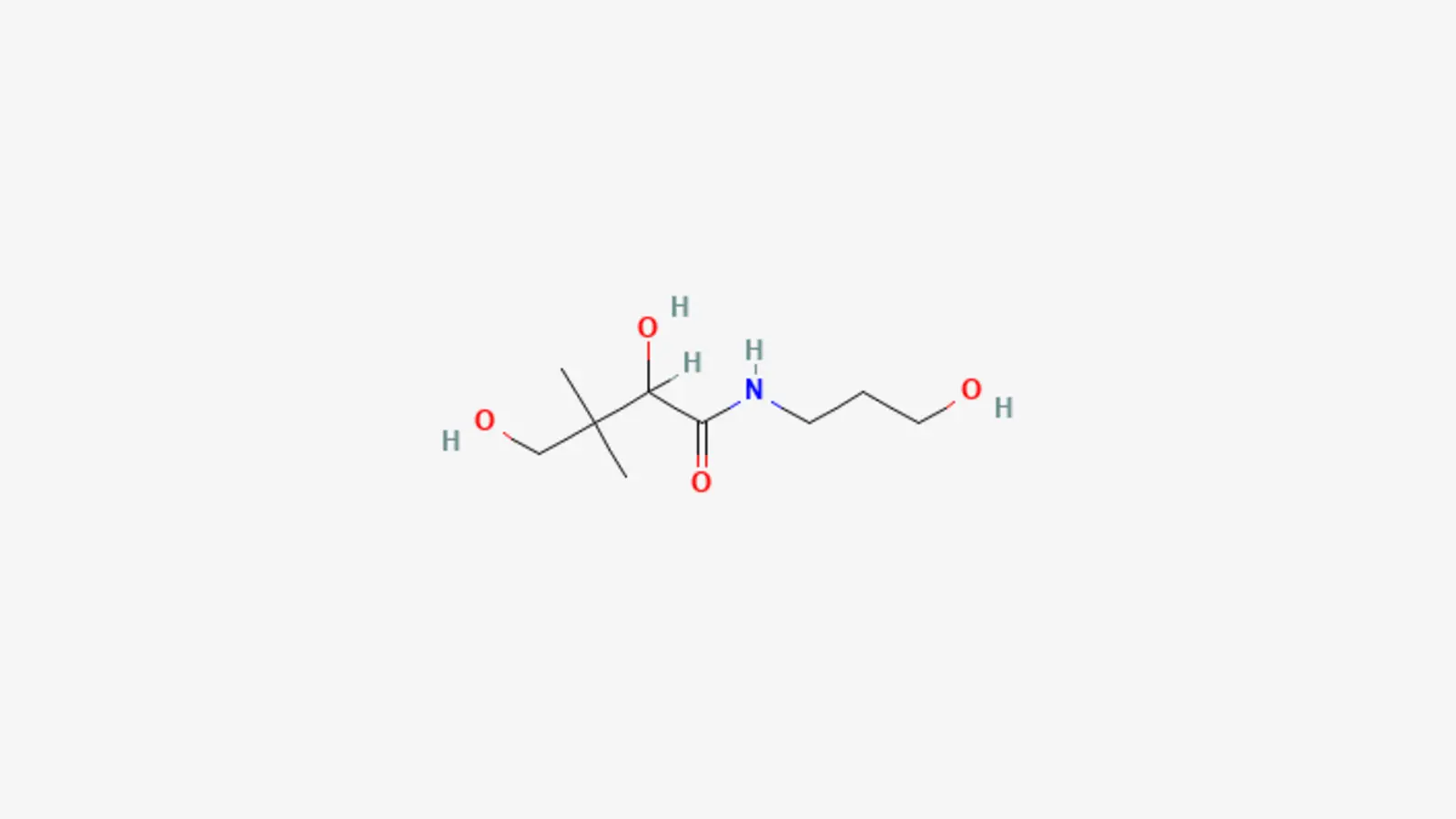
What is Panthenol: Scientific Mechanisms and Clinical Evidence
Read article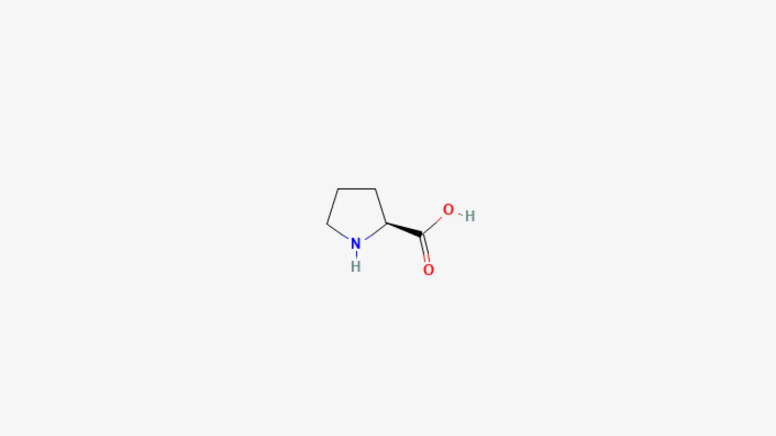
What is Proline: Scientific Mechanisms and Clinical Evidence
Read article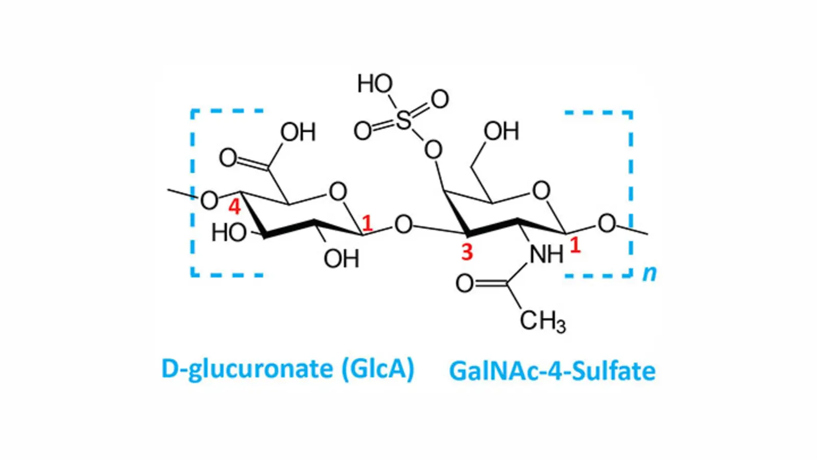
What is Proteoglycan: The Scientific Mechanisms and Clinical Evidence
Read article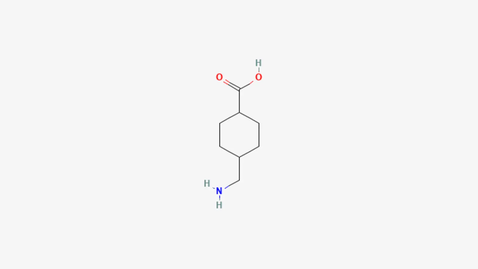
What is Tranexamic Acid: The Scientific Mechanisms and Clinical Evidence
Read article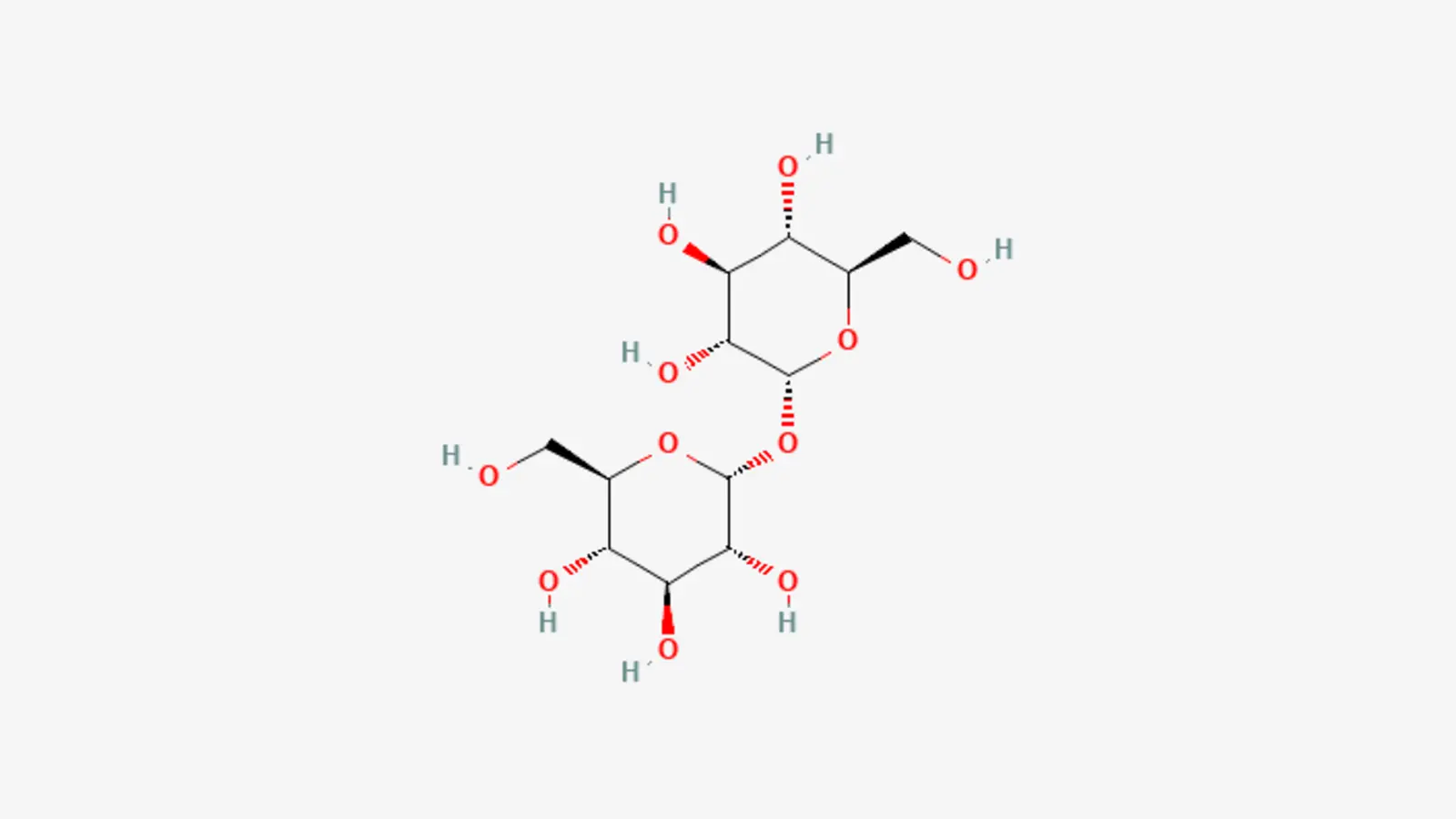
What is Trehalose: Scientific Mechanisms and Clinical Evidence
Read article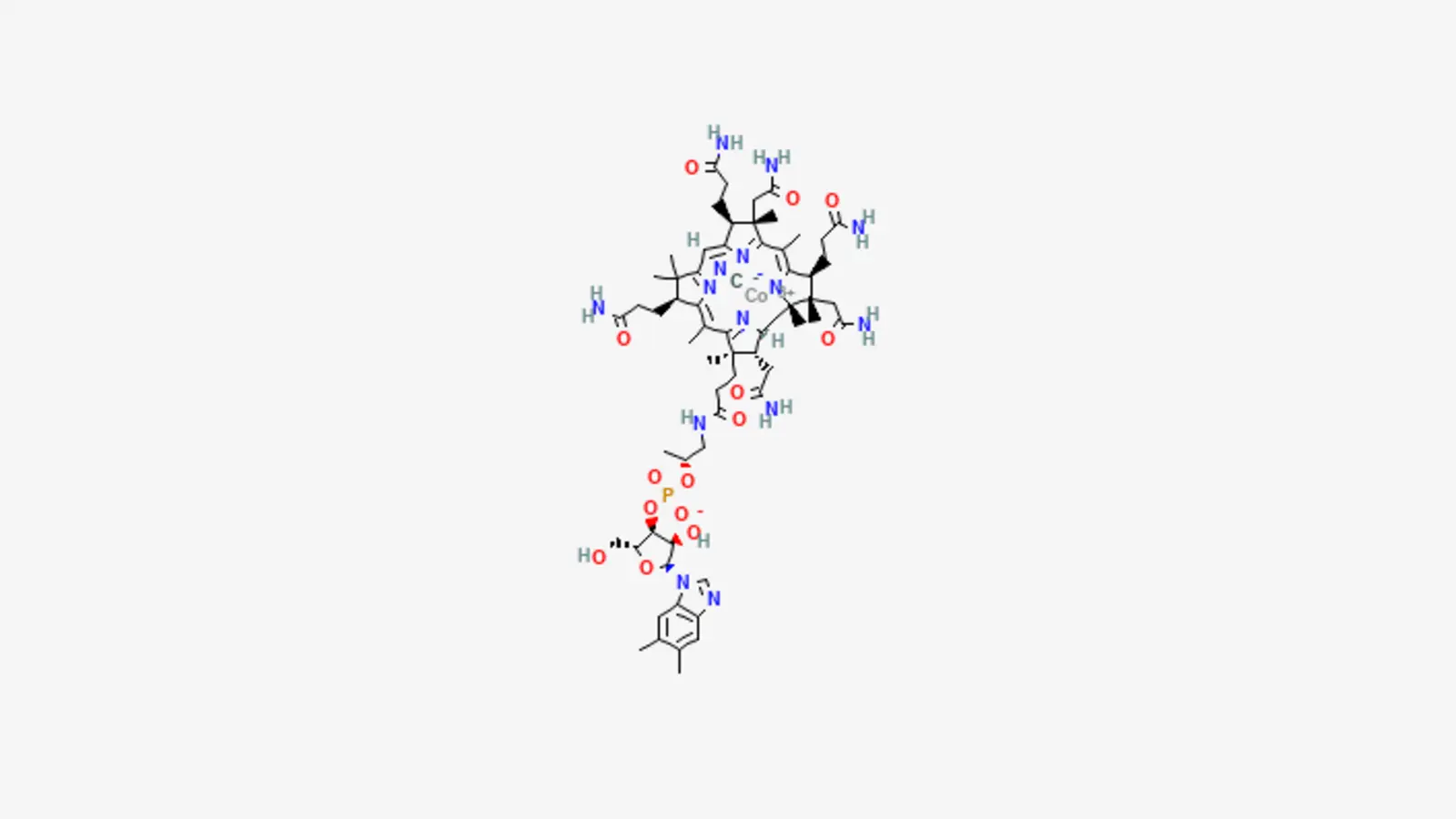
What is Vitamin B12: The Scientific Mechanisms and Clinical Evidence
Read article
Cellular Reprogramming: How Exosomes and Paracrine Signaling Reset the Regenerative Clock in Aging Skin
Read article
Deep Dermal Bioremodeling: How Polynucleotides (PN/PDRN) Activate A2A Receptors to Repair Atrophic Tissue
Read article
Epidermal Lipid Reconstruction: How Ceramides and Phytosterols Repair an Over-Exfoliated Skin Barrier
Read article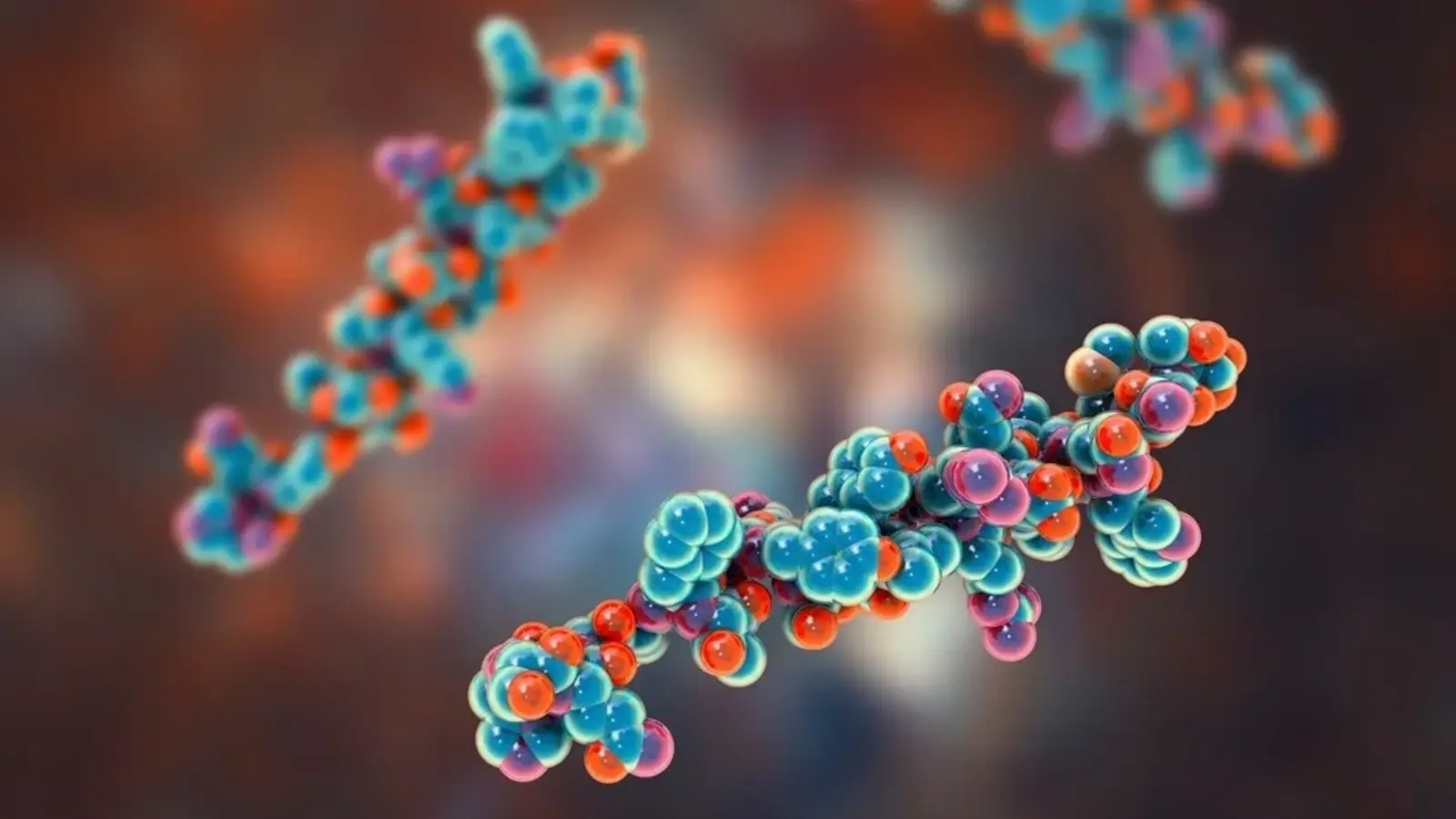
Extracellular Matrix Restructuring: How GHK-Cu and Acetyl Tetrapeptide-2 Target Horizontal Neck Creases
Read article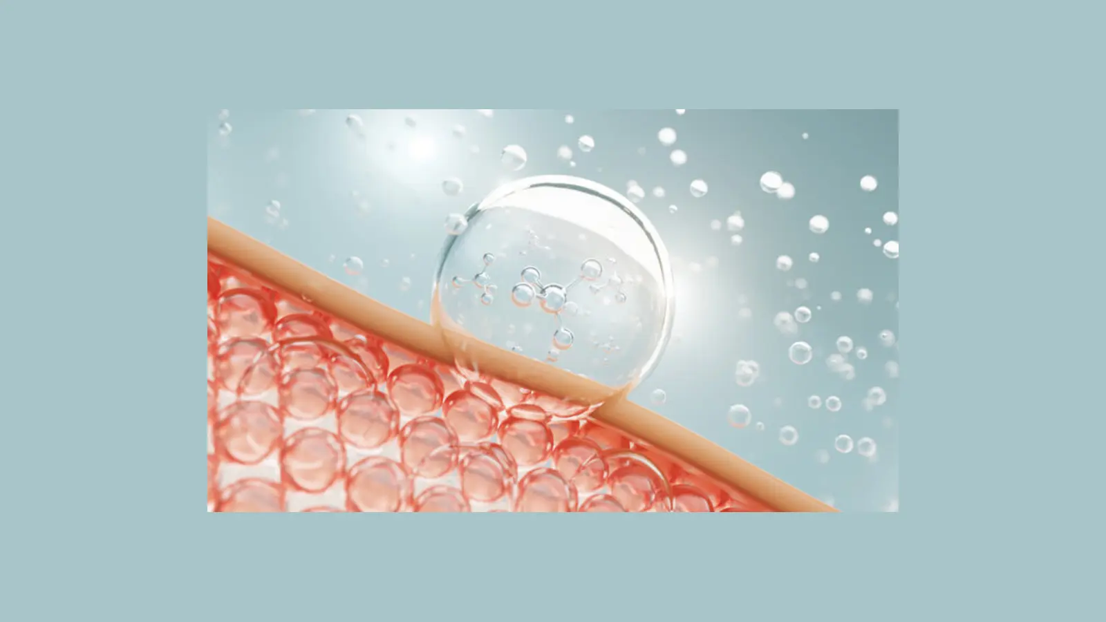
Mitochondrial Resuscitation: How Coenzyme Q10 and NADH Recharge Sluggish, ATP-Deprived Skin Cells
Read article
Molecular Moisture Locks: Reversing Deep Dehydration and Reclaiming “Glass Skin” Glow
Read article
Neuromuscular Relaxation: How SNAP-8 Peptide Softens Perioral Lines and Restores Lip Border Definition
Read article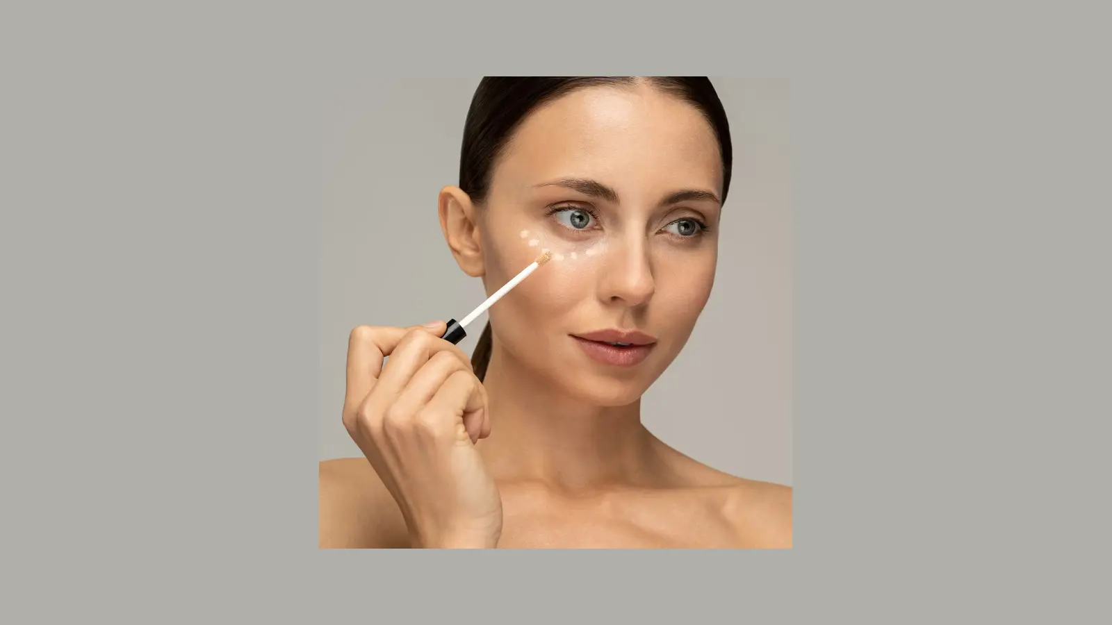
Surface Hydrophobicity and Sebaceous Control: How Silica Silylate and Zinc Glycinate Eliminate Midday Shine
Read article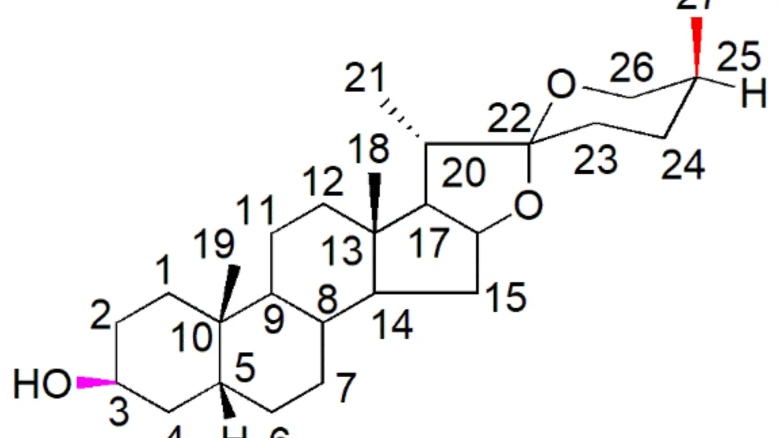
Targeted Adipogenesis: How Sarsasapogenin Reverses Facial Fat Pad Atrophy and Mid-Face Hollowing
Read article
Targeting Deep Dermal Melanin: Biological Pathways of Cysteamine HCl and Oligopeptide-68 in Recalcitrant Melasma
Read article
Calming the Brain-Skin Axis: How Palmitoyl Tripeptide-8 Neutralizes Neorogenic Inflammation and Stress-Flushing
Read article
Depuffing ‘Cortisol Face’: How Topical Adaptogens Neutralize Stress Swelling and Dullness
Read article
Fading ‘Red Marks’ Fast: How Tranexamic Acid and Potassium Azeloyl Diglycinate Clear Post-Acne Erythema (PIE)
Read article
Intravascular Fortification: How Troxerutin and Escin Seal Capillary leaks to Clear Diffuse Facial Flushing
Read article
Oxygen Resuscitation: How Perflurodecalin Recharges Hypoxic, Asphyxiated Dermal Tissue
Read article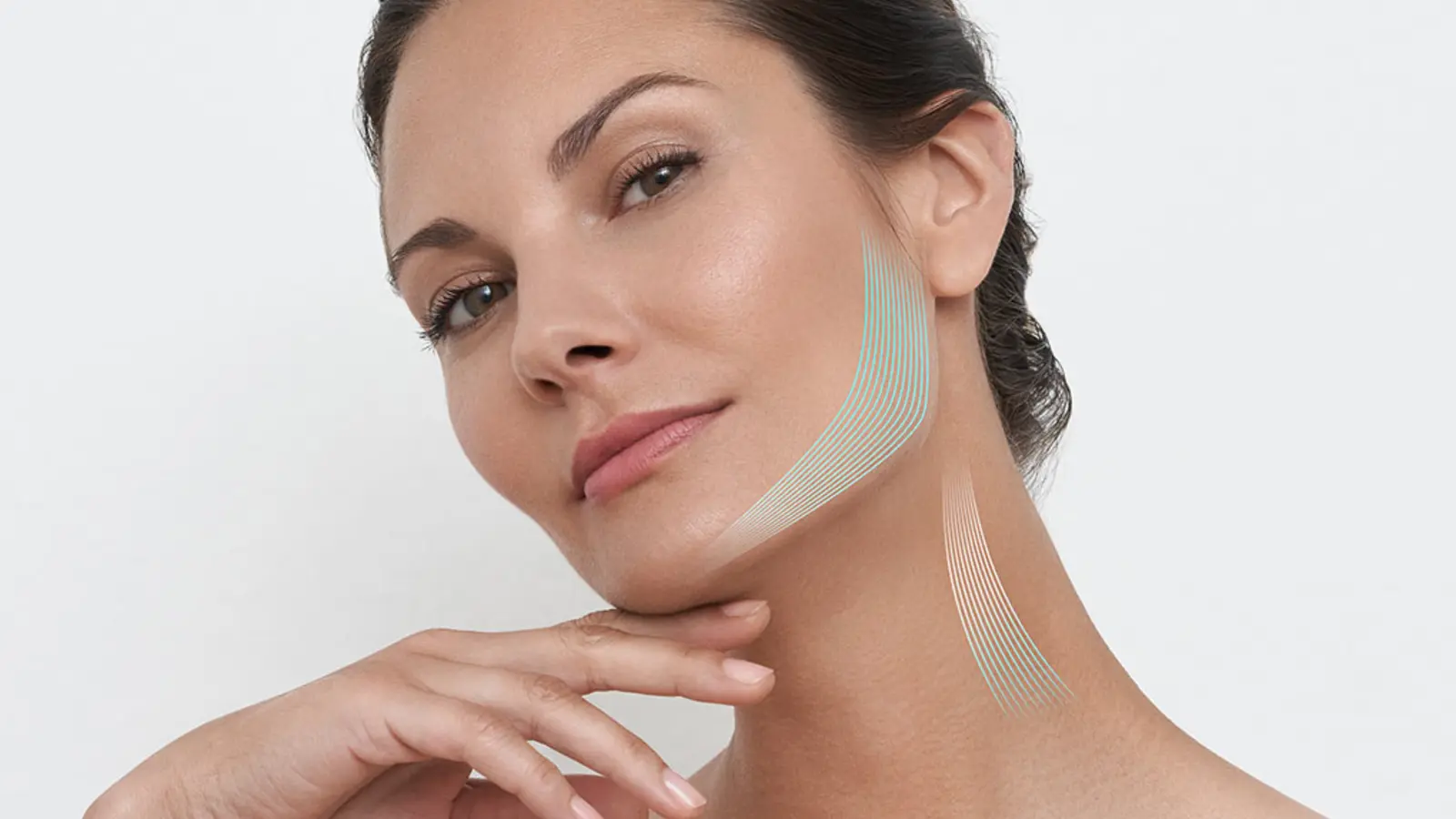
Re-Sharpening the Jawline: How DMAE Tightens ‘Tech-Neck’ Laxity
Read article
Reversing ‘Sugar Sag’: How Carnosine and Alpha-Lipoic Acid Un-Crosslink Glycated Dermal Tissue
Read article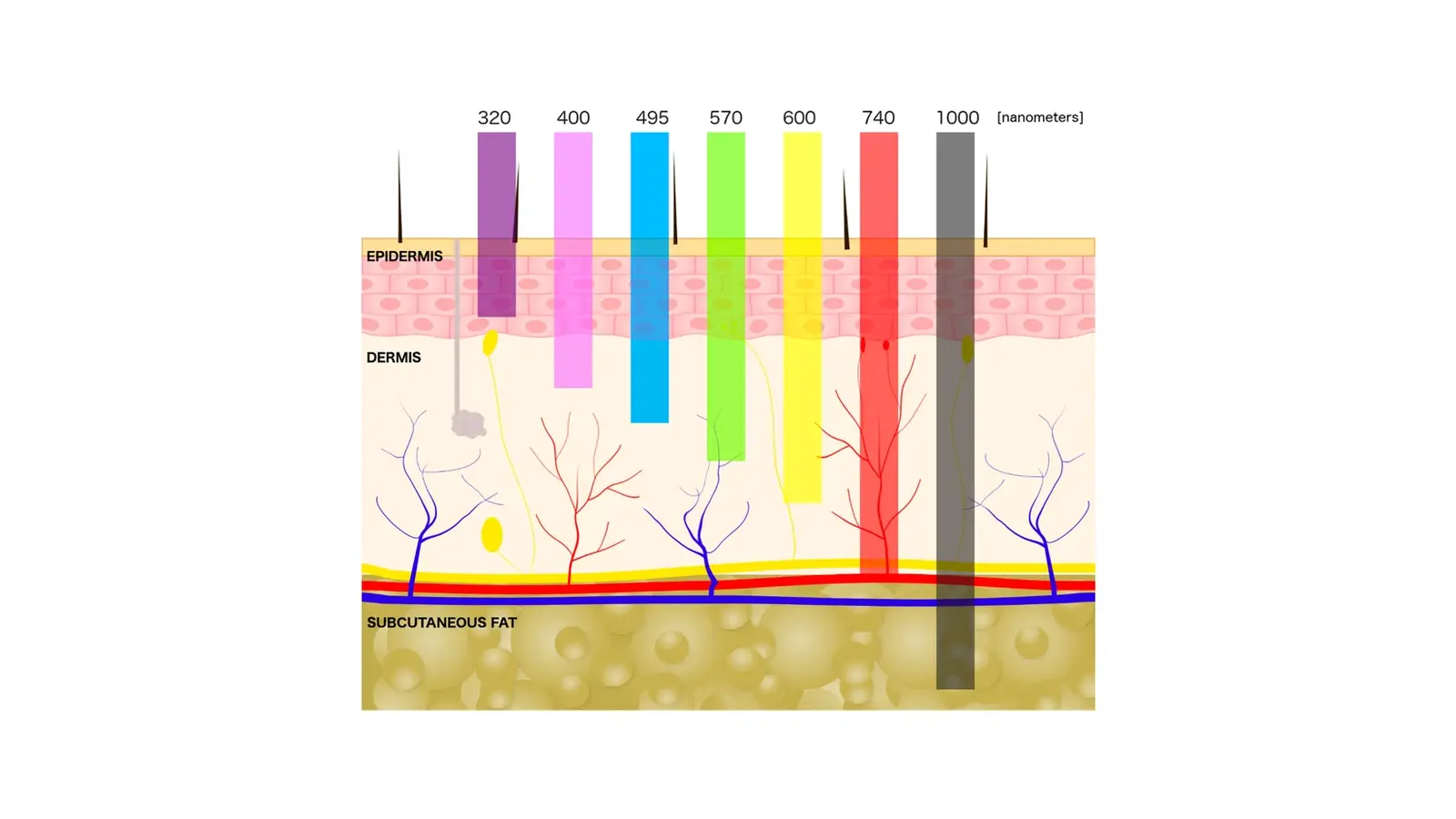
Screening Out Pigment: How Luitein and Ectoin Shield Skin from Digital Blue Light
Read article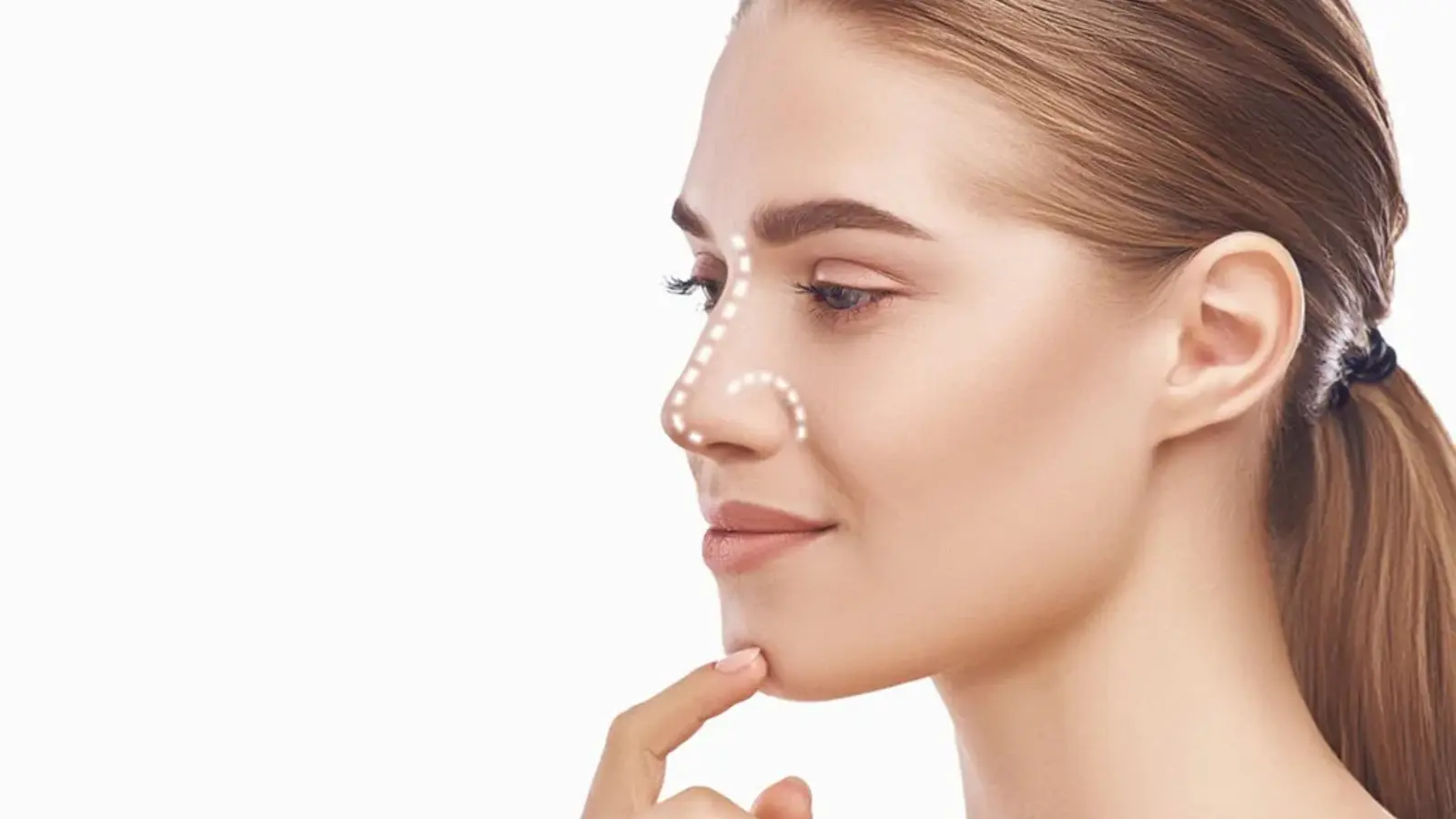
Smooth Out ‘Strawberry Nose’: How Lipo-Hydroxy Acid De-Clogs Sebaceous Filaments Without Peeling
Read article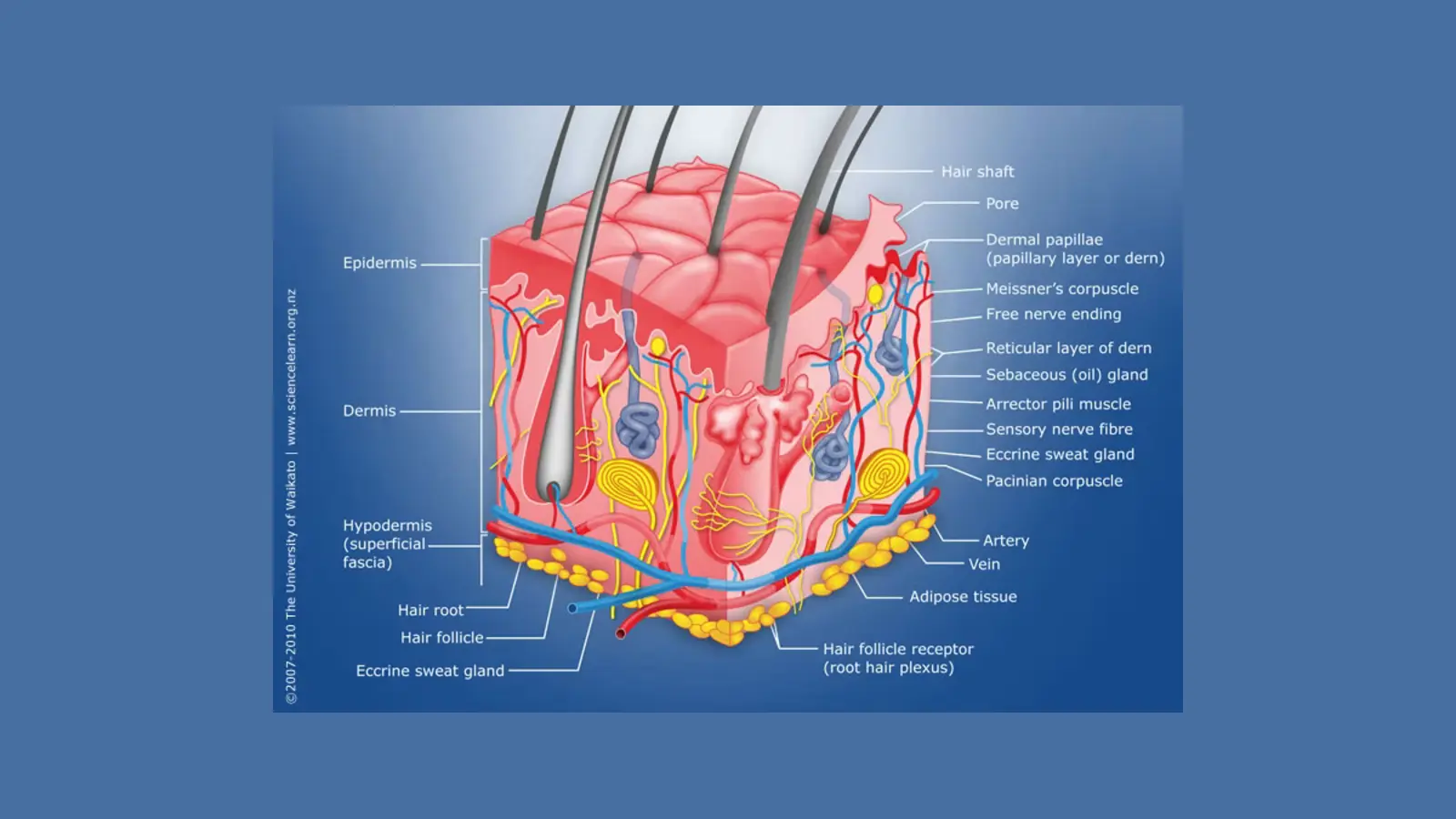
The Senolytic Shift: How Fisetin and Quercetin Clear ‘Zombie Cells’ to Re-Energize Dermal Tissue
Read article
Beyond Hydroquinoe: How Cysteamine HCl Masterminds Safe, Long-Term Pigment Correction
Read article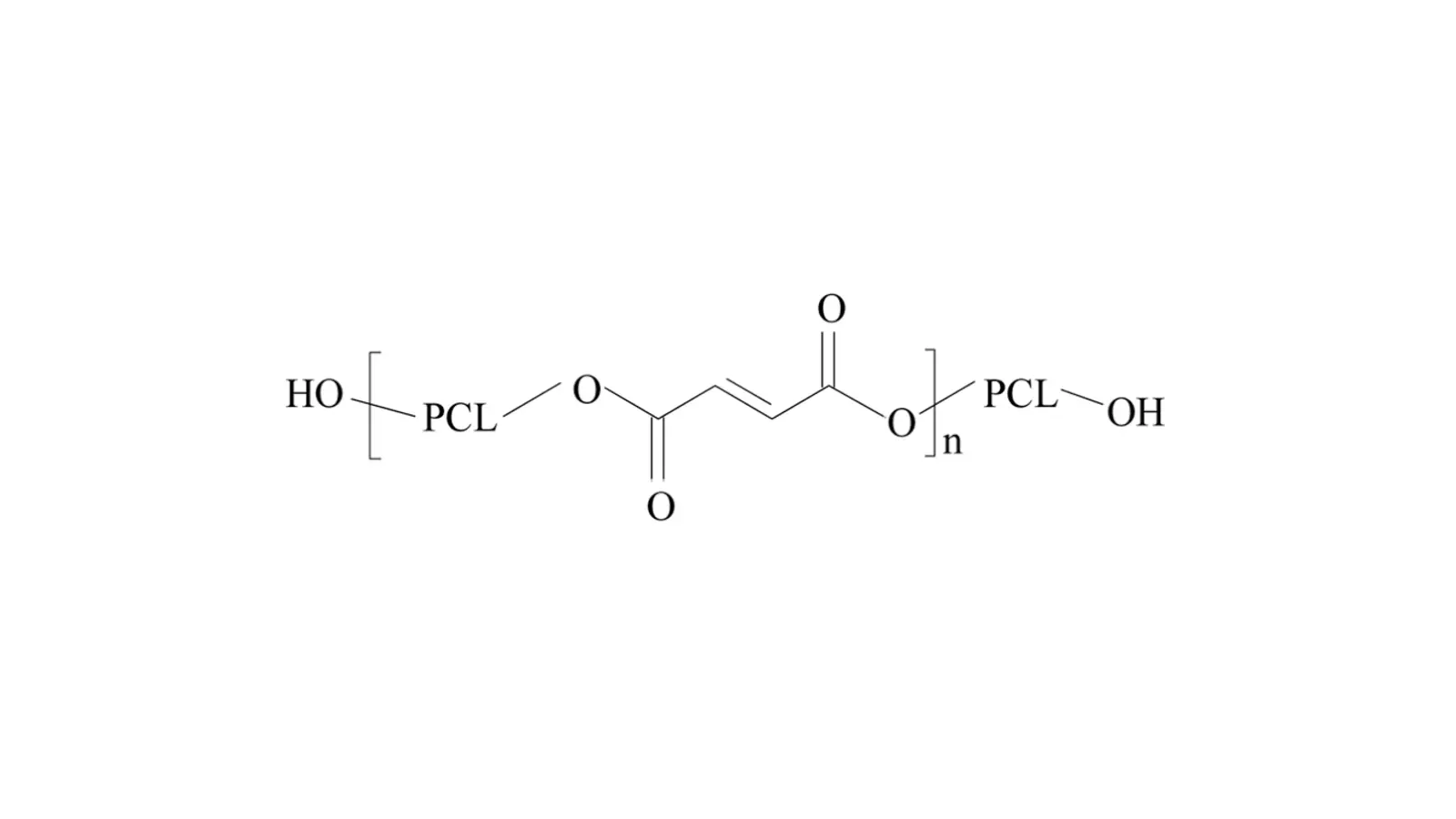
Beyond Volume Loss: How Polycaprolactone (PCL) Rebuilds the Mid-Face Dermal Matrix
Read article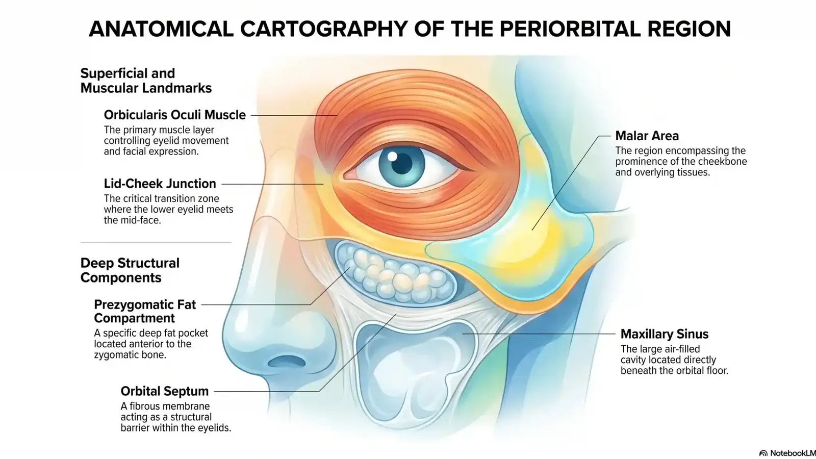
De-Puffing Science: How Caffeine and EGCG Flush Stagnant Periorbital Fluid
Read article
Fixing ‘Ozempic Face’: How Advanced Peptides Re-Densify Skin After Rapid Weight Loss
Read article
Precision Micro-Filling: How 3D-Network HA Smooths Periorial Verticle Creases
Read article
Re-Anchoring the Epidermis: How rhCollagen Restores the Dermal-Epidermal Junction
Read article
Reversing Cellular Solar Damage: How Photolyase DNA Repair Enzymes Restore Photoaged Skin
Read article
The Needle-Free Brow Lift: How Argireline Amplified Relaxes Periorbital Tension
Read article
The Pore-Refining Power Couple: Inside the Protocol for Matte, Filter-Smooth Skin
Read article
The Ultimate Shield: How Ectoin Calms Extreme Post-Treatment Skin Sensitivity
Read article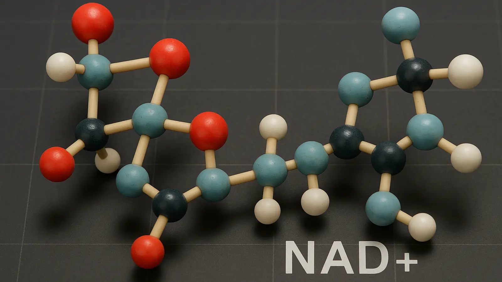
Cellular Recharging: How NAD+ Turns Back the Clock on Sluggish, Stressed Skin
Read article
Smooth the Creases: How Synthetic Snail Glycoprotiens are Conquering ‘Tech-Neck’ and Texture
Read article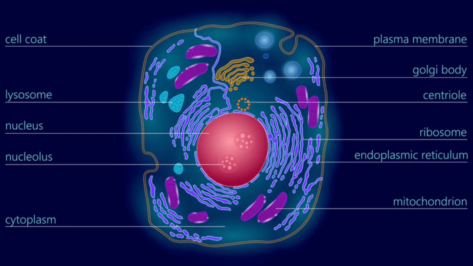
The Laser Co-Therapy Secret: How Exosomes cut your Post-Treatment Downtime in Half
Read article
The Needle-Free Fill: How Polyglutamic Acid Re-Inflates Deflated Skin
Read article
The ‘Glass Skin’ Shortcut: Why Non-Crosslinked HA and Glycerol Are Winning the Aesthetic Lounge
Read article
Beyond Tear Through Fillers: How Sodium Succinate Rejuvenates the Under-Eye Cellular Matrix
Read article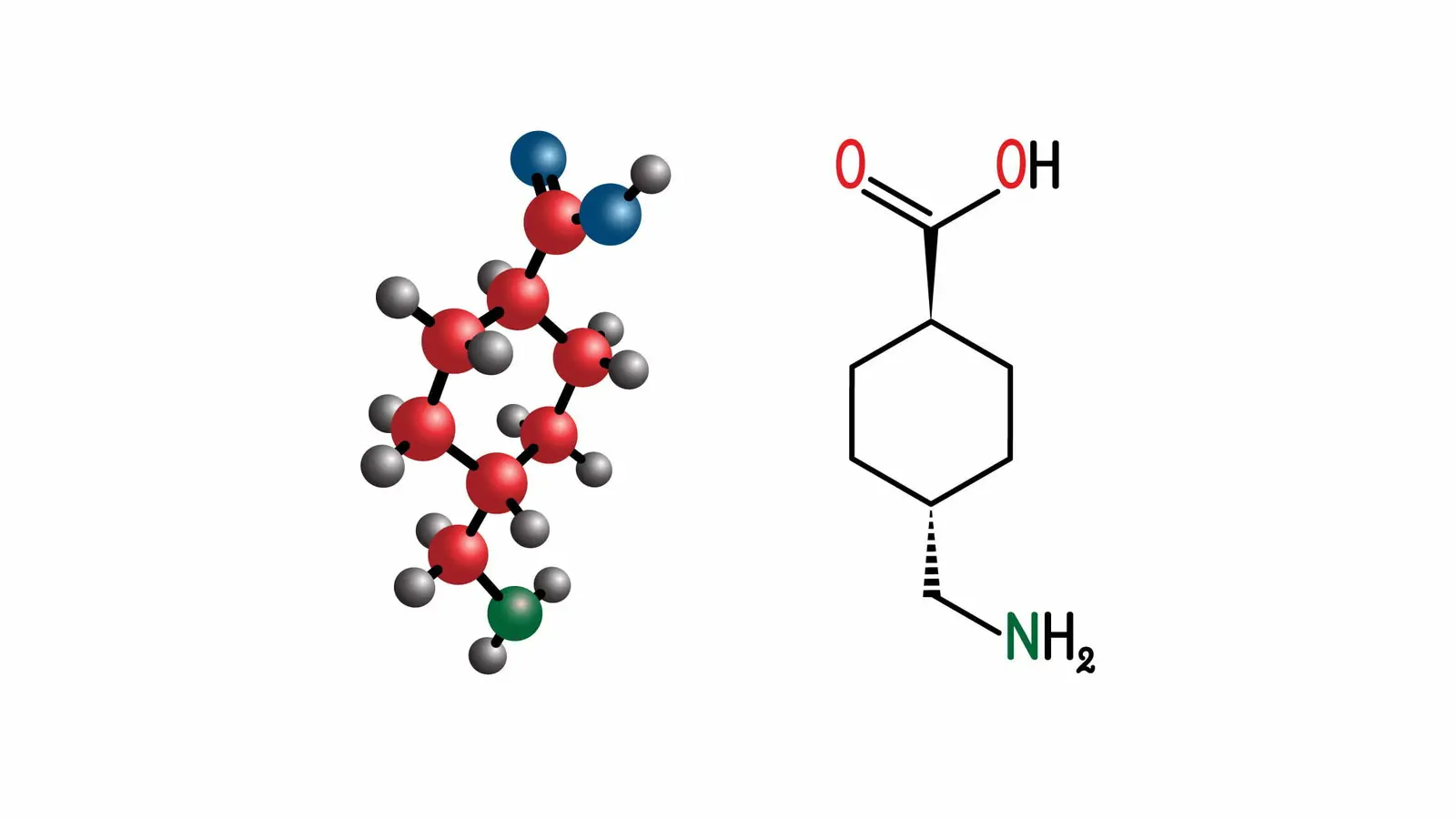
Shifting the Paradigm on Pigment: The New Protocol for Post-Inflammatory Erythema
Read article
The Neurotoxin Alternative: Deep Wrinkle Remodeling with GHK-Cu Copper Peptides
Read article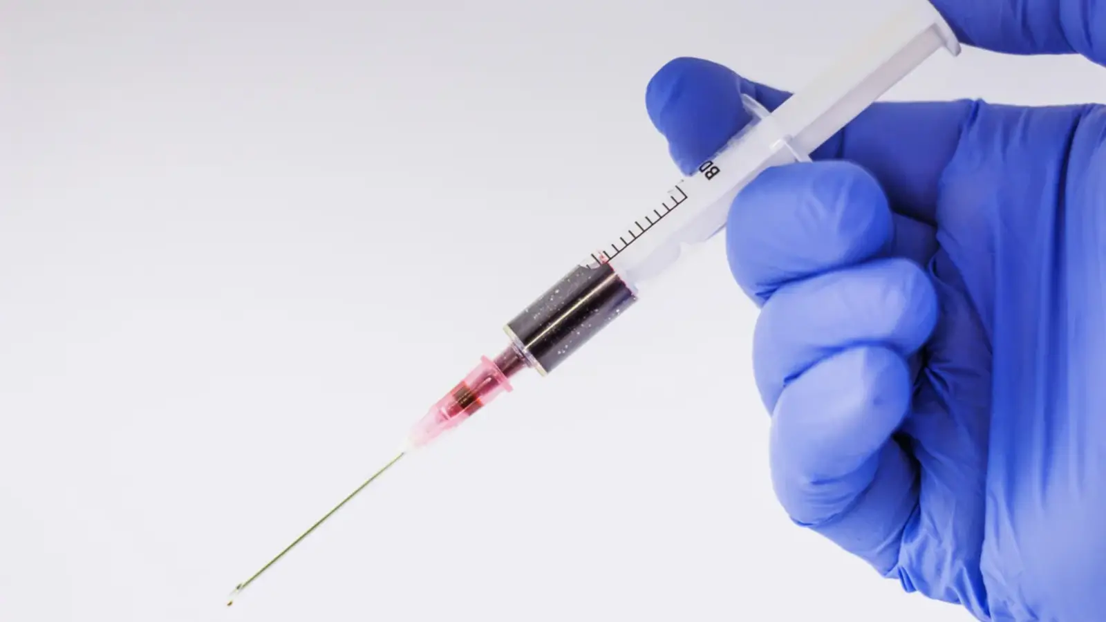
Non-Surgical Jawline Contouring: What to Expect from Lipolytic Injections
Read article
The Science of Biostimulation: How PLLA Rebuilds Lost Facial Architecture
Read articleNo articles match your search.